Case 94: Going the Extra Mile in Anterior Esthetic Treatment

A 32-year-old woman presented with an initial concern about the appearance of her smile and a fistula at tooth no 8: “I’m very worried about my smile. I have a fistula and need to know about implants. I wish my teeth were more properly shaped—actually, I wish they were perfect.” The patient received orthodontic therapy in 7th grade. While in high school she was elbowed in the mouth during a basketball game and sustained an injury that went untreated for 2 years, at which point she underwent root canal therapy on teeth nos. 8 and 9 followed by a tissue graft. At age 20 she had veneers placed on those teeth. Four years later she had root exposures and new veneers were placed on teeth nos. 7, 8, 9, and 10. In 2011, the veneers on teeth nos. 8 and 9 were replaced with crowns, and her dentist promised that this would be “longer lasting.” A few months later, a fistula developed at tooth no 8. Finally, she reported that in 2012, she awakened in an open locked condition.

Other Concerns

  • Wants a wider smile.
  • Wants wider centrals and longer laterals.
  • Dislikes the position of tooth no 27.
  • Dislikes all the Class V composites and feels they make her teeth look long and unnatural.

Medical History

  • Current medications: Zyrtec-D, Beyaz (oral contraceptive).
  • Known drug allergies: None (environmental only).
  • Smoking status: Nonsmoker.
  • Alcohol status: Drinks socially.
  • ASA physical status classification: ASA I.

Diagnostic Findings

Extraoral/Facial

  • Within normal limits (WNL).

TMJ/Mandibular Range of Motion/Muscles of Mastication and Facial Expression

  • TMJ evaluation: Grating on right side.
  • History of TMD and utilization of appliances to relieve pain.
  • Range of mandibular motion WNL. No deviation on opening/closing.
  • Pain on palpation of the right and left mid-portion masseter muscles.
  • Mild pain on palpation of the right and left lateral pterygoid muscles.

Intraoral Findings

Dental

  • Missing teeth nos. 1, 16, 17, and 32.
  • Oral cancer examination WNL.
  • Dental caries: None.
  • Salivary flow: Normal.
  • Overcontoured restorations on teeth nos. 7, 10, and 11.
  • Mandibular midline 1 mm to the right of maxillary midline.

Soft Tissue/Periodontal

  • General level of gingival inflammation is mild with good plaque control.
  • Biologic width violation teeth nos. 8 and 9.
  • Purulence/bleeding upon probing tooth no. 8.
  • Teeth nos. 8 and 9 have mobility of 1.
  • Gingival recession teeth nos. 2, 3, 5, 6, 11–13, 21–23.
  • Mucogingival abnormalities at teeth nos. 11, 20–22, 27 (lack of attached gingiva).

FMX 2013

Occlusal Notes

  • Maxillomandibular relationship is Class I.
  • Overbite 1 mm.
  • Overjet 1 mm.
  • Angle molar classification: Class I right and left sides.
  • Canine relationship: Class I right and left sides.
  • Fremitus: Teeth nos. 8 and 9.
  • Centric relation/initial point of contact on teeth nos. 15/18.
  • 1 mm vertical slide from CR to CO.
  • Altered Curve of Spee: Right and left sides.
  • Balancing interferences on teeth nos. 3/31, 13/19, 14/19, and 15/18.
  • Right working guidance on teeth nos. 15/18.
  • Protrusive guidance on posterior teeth.
Initial maximum intercuspal position.
Initial protrusion.
Pretreatment right lateral view.
Pretreatment left lateral view.
Pretreatment maxillary occlusal view.
Pretreatment mandibular occlusal view.

CBCT Imaging Review

Relevant to Anterior Maxilla

  • Lateral pathology on teeth nos. 8 and 9.
  • Invasive resorption on tooth no. 9.
  • Lack of palatal bone on teeth nos. 8 and 9.
  • Loss of proximal crestal bone between teeth nos. 7/8, 8/9, and 9/10.
Initial maxillary anterior PAs.

Diagnosis

  • Localized recession-based attachment loss with mild to moderate loss of periodontal support teeth nos. 2, 3, 5, 11–13, 21–23.
  • Vertical root fracture tooth no. 8.
  • Invasive cervical resorption tooth no. 9.
  • Mucogingival abnormalities teeth nos. 11, 20–22, 27.
  • Gingival inflammation secondary to biologic width violation teeth nos. 8 and 9.
  • Right side anterior open bite possibly secondary to tongue posturing habit.
  • Moderate degenerative changes of the right condyle.

Prognosis

  • Poor for teeth nos. 8 and 9.
  • Good for all other teeth.

Summary of Concerns

  • Our immediate concern is how to deal with failing teeth nos. 8 and 9.

After emergent management of infection:

  • How do we handle the soft tissue and esthetics given the bone loss both lingually and interproximally at sites nos. 7, 8, 9, and 10?
  • How do we handle the constricted arch forms and spacing requirements for ideal esthetics?
  • How do we handle the right-side anterior open bite?
  • Will correcting the patient’s occlusion help resolve her longstanding TMD/myalgia issues?

Proposed Treatment Plan

Phase I: Address Failing Teeth

  • Extract teeth nos. 8 and 9 and socket graft with rhBMP-2 and cancellous allograft.
  • Fabricate a provisional bridge across teeth nos. 7–10 with ovate pontics to maintain soft tissue form.

Phase II: Diagnostic Work-up

  • Obtain a complete set of radiographs, a CBCT scan, clinical and periodontal charting, and diagnostic models mounted with facebow and CR bite records.
  • Formulate a comprehensive treatment plan based on consultation between restorative, periodontal, and orthodontic specialists.

Phase III: Periodontics/Orthodontics

  • Perform surgically facilitated orthodontic therapy (SFOT) to allow for expansion orthodontia.
  • Complete guided bone regeneration procedure in the anterior maxilla for development of future implant sites nos. 8 and 9 at the time of SFOT surgery.
  • Complete connective tissue grafting to gain attached tissue and root coverage at sites nos. 21, 22, and 27.

Phase IV: Implant Placement

  • Re-image the maxillofacial complex following SFOT and site development at teeth nos. 8 and 9. Determine final plans post bone reconstruction.
  • Complete guided implant placement at sites nos. 8 and 9 along with contour bone grafting and/or connective tissue grafting, as needed.

Phase V: Second Stage Implant Surgery

  • Expose implants.

Phase VI: Provisional Restorative Treatment

  • Provisionalize implants nos. 8 and 9 to groom the soft tissues for ideal esthetics.
  • Provisionalize any other teeth as necessary for ideal esthetics.

Phase VII: Definitive Restorative Treatment

  • Place definitive restorations on teeth nos. 7 and 10 and on implants nos. 8 and 9.
  • Place any other definitive restorations.

Phase VIII: Occlusal Splint Therapy

  • Undertake splint therapy if needed after orthodontic occlusal correction.
  • Schedule regular periodontal, restorative, occlusal, and implant maintenance.
I/C=implant supported crown; C=crown © Seattle Study Club Journal

Active Clinical Treatment

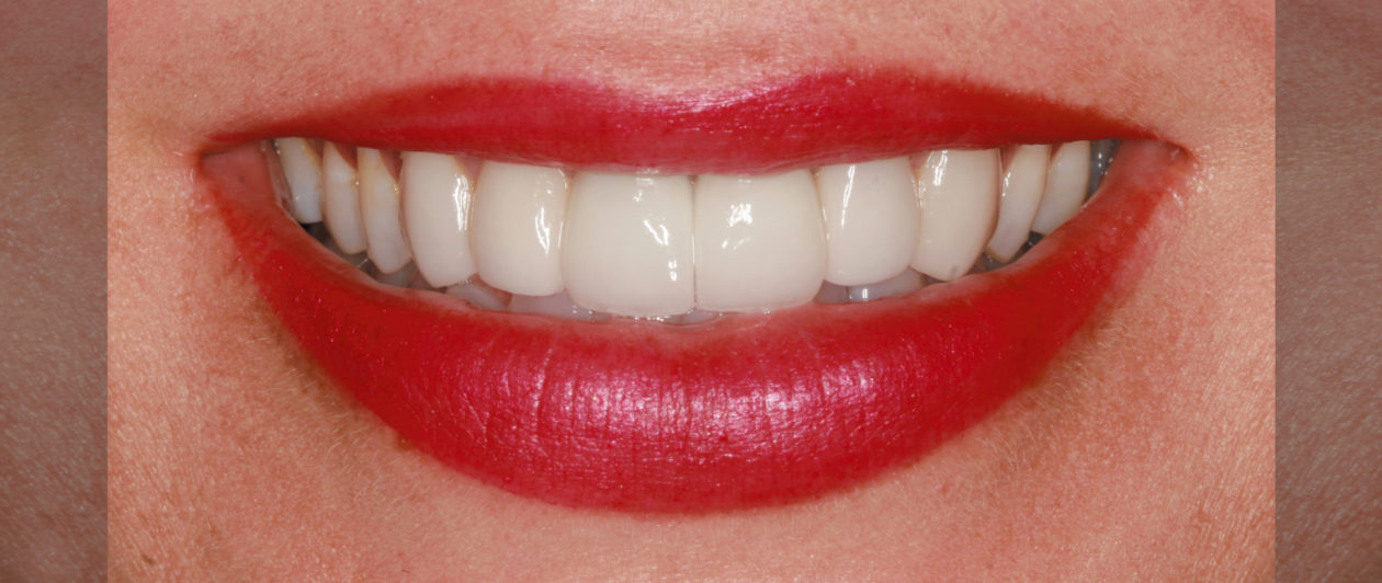
Review of Treatment Goals

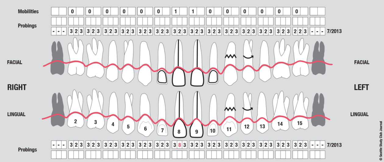
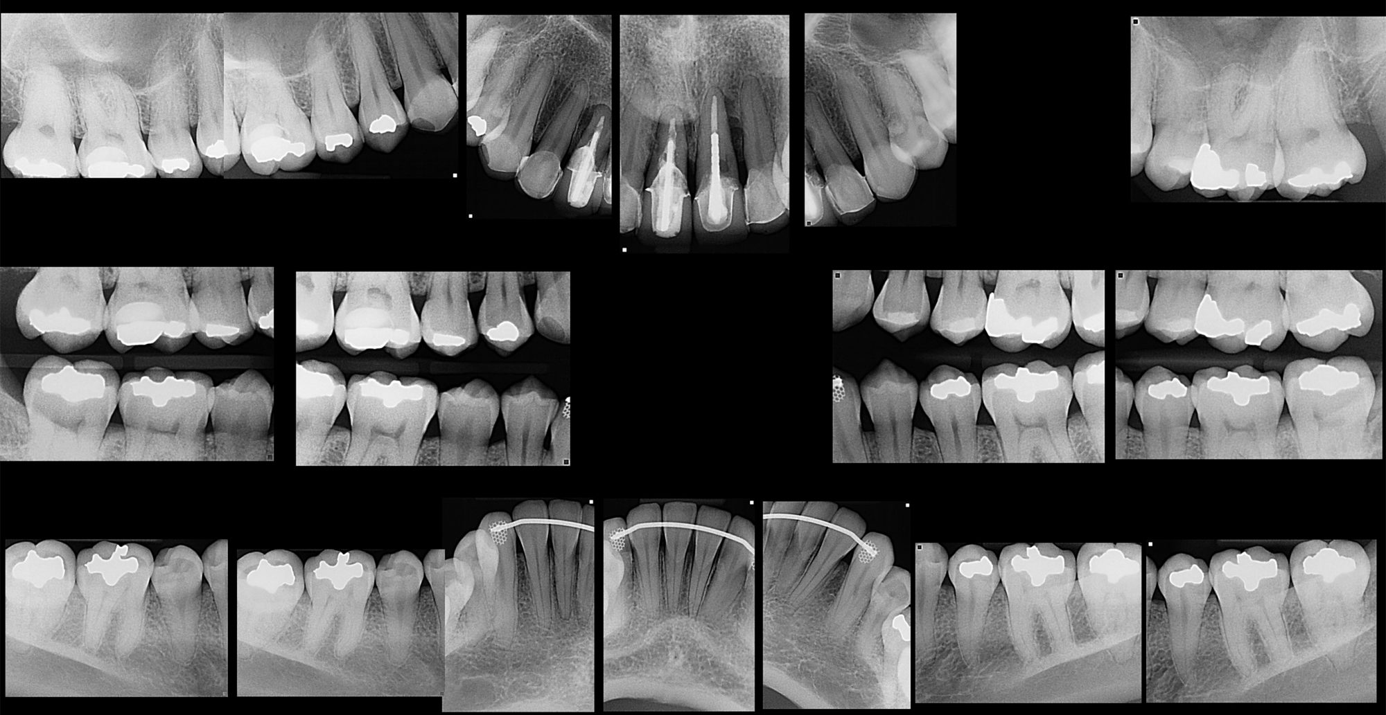
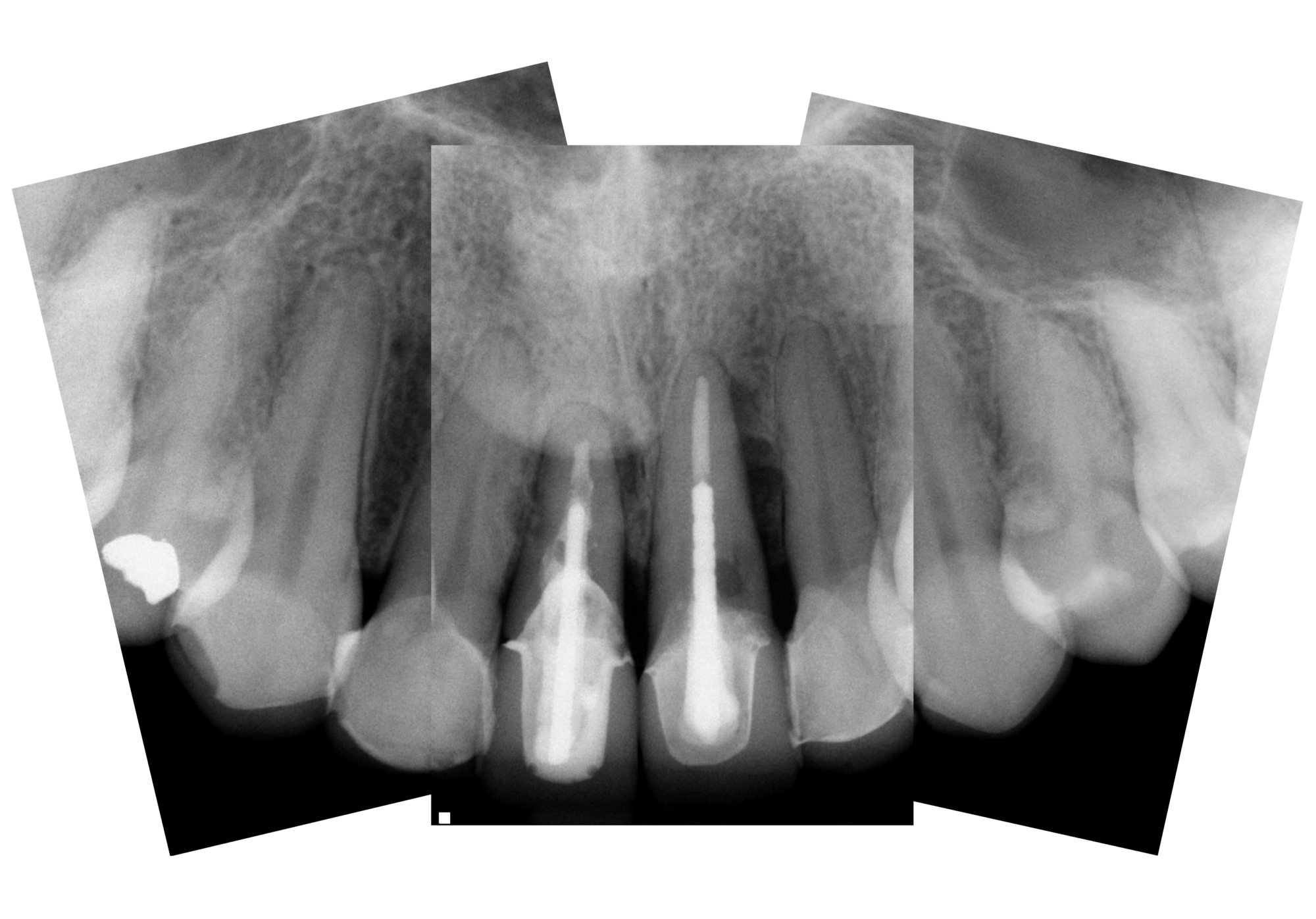
The patient presented with an initial concern about the appearance of her smile and a fistula at tooth no 8. She completed orthodontic therapy in 7th grade but was elbowed in the mouth a few years later and sustained an injury that went untreated for 2 years and ultimately required root canal therapy on teeth nos. 8 and 9, followed by a tissue graft. At age 20 she had veneers placed on those teeth. Four years later she had root exposures, and new veneers were placed on teeth nos. 7–10. In 2011, the veneers on teeth nos. 8 and 9 were replaced with crowns. In 2012, she awakened one day in an open locked condition.

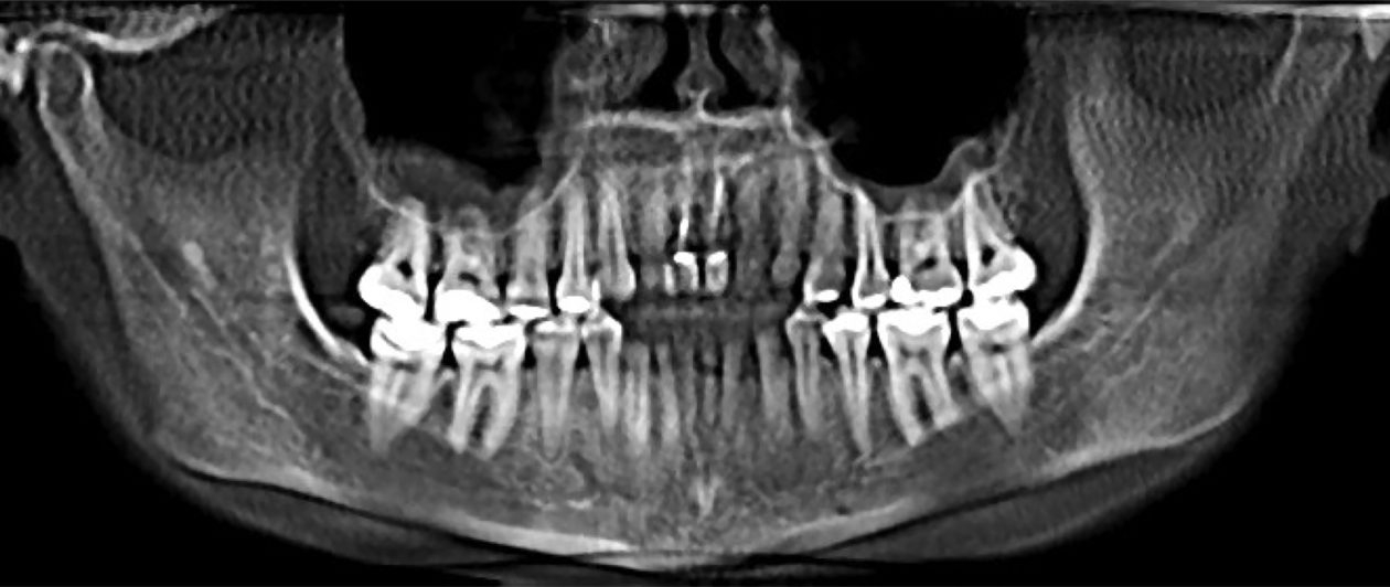
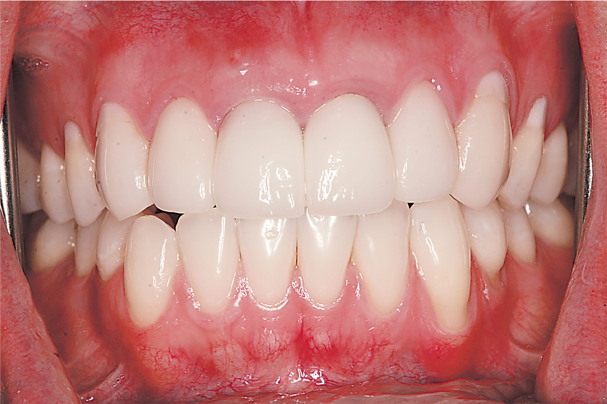
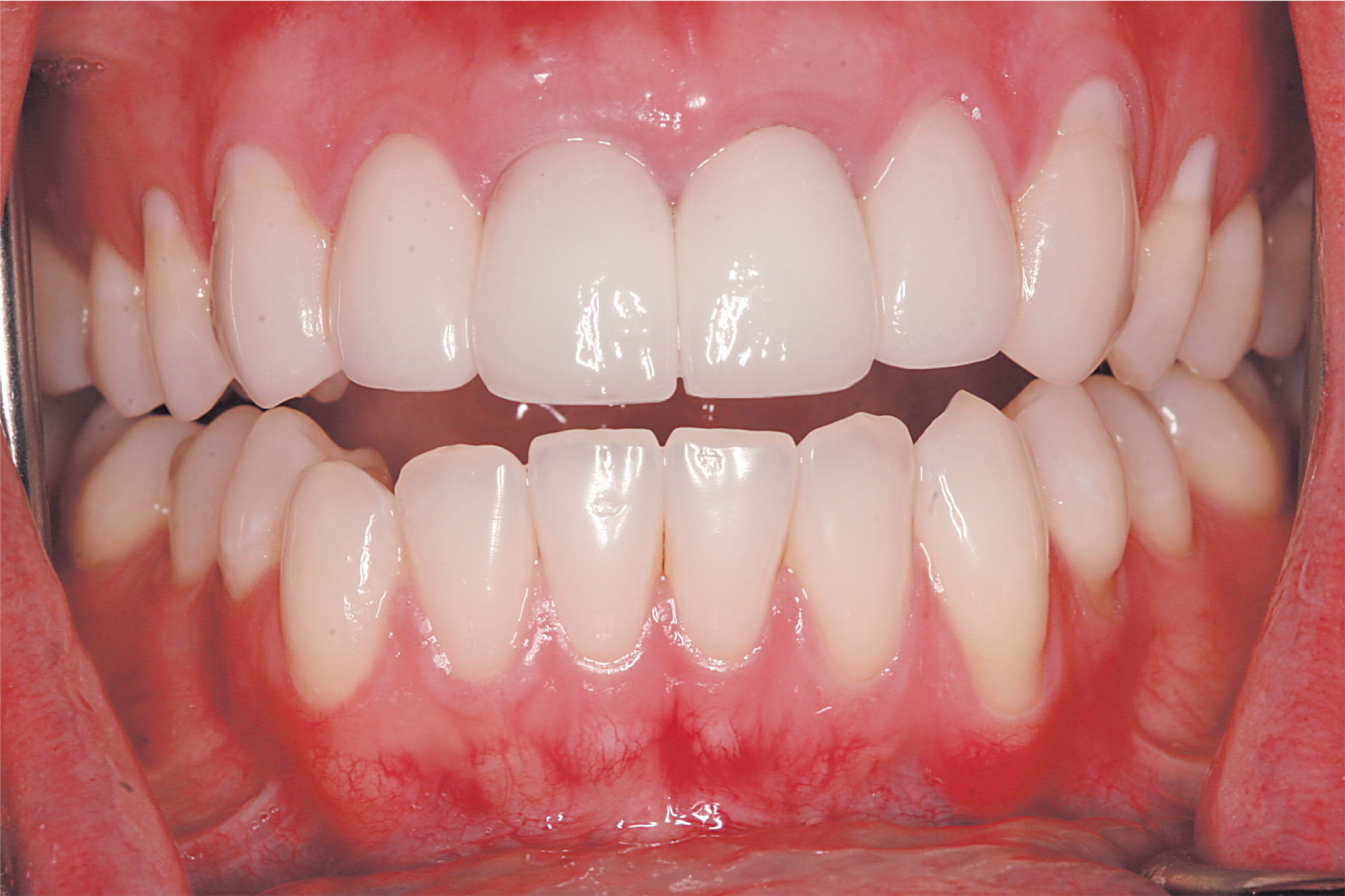
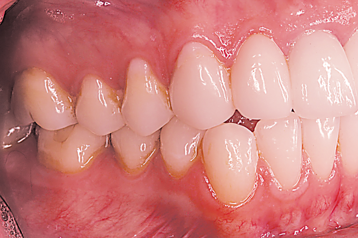
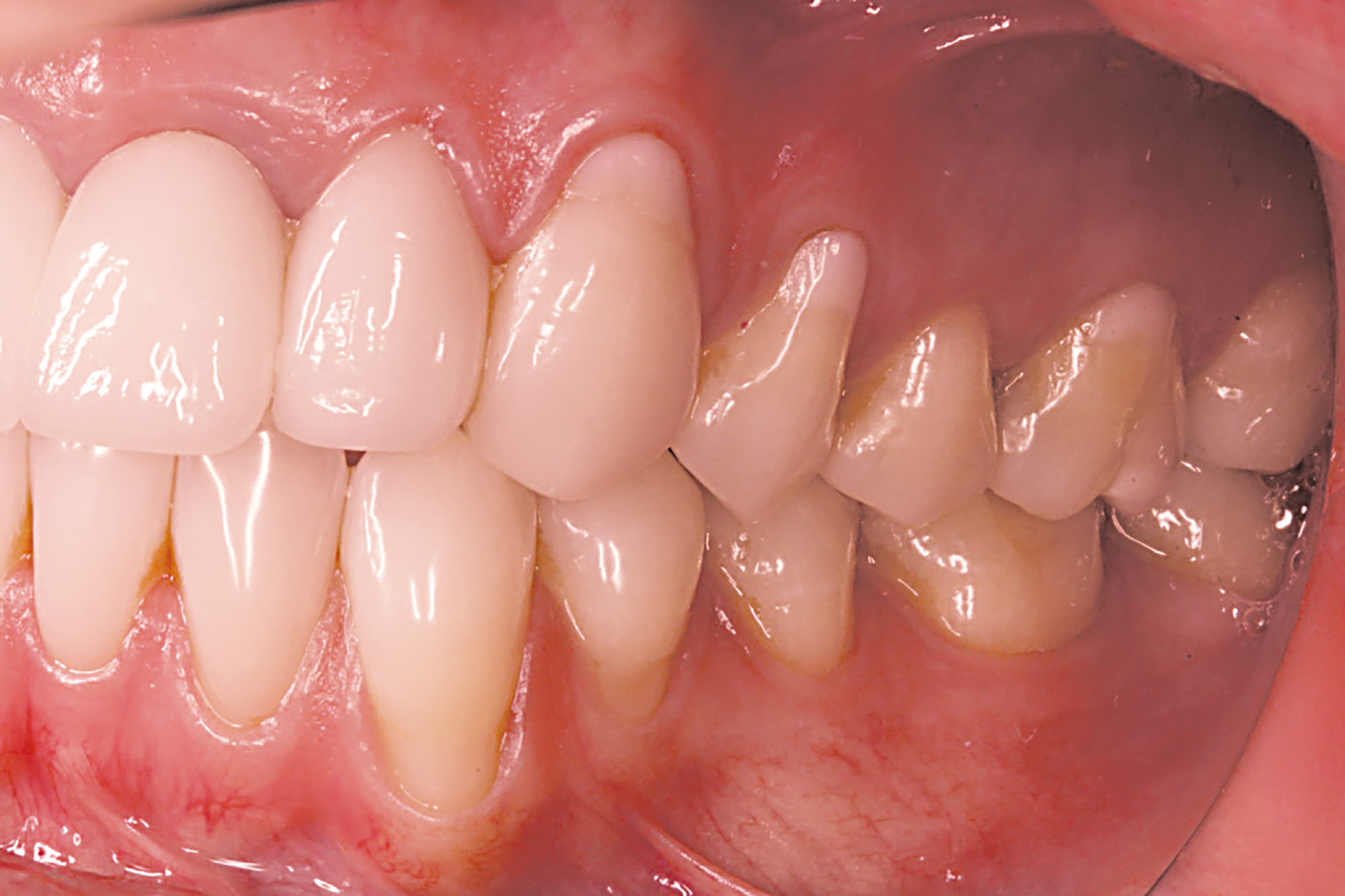
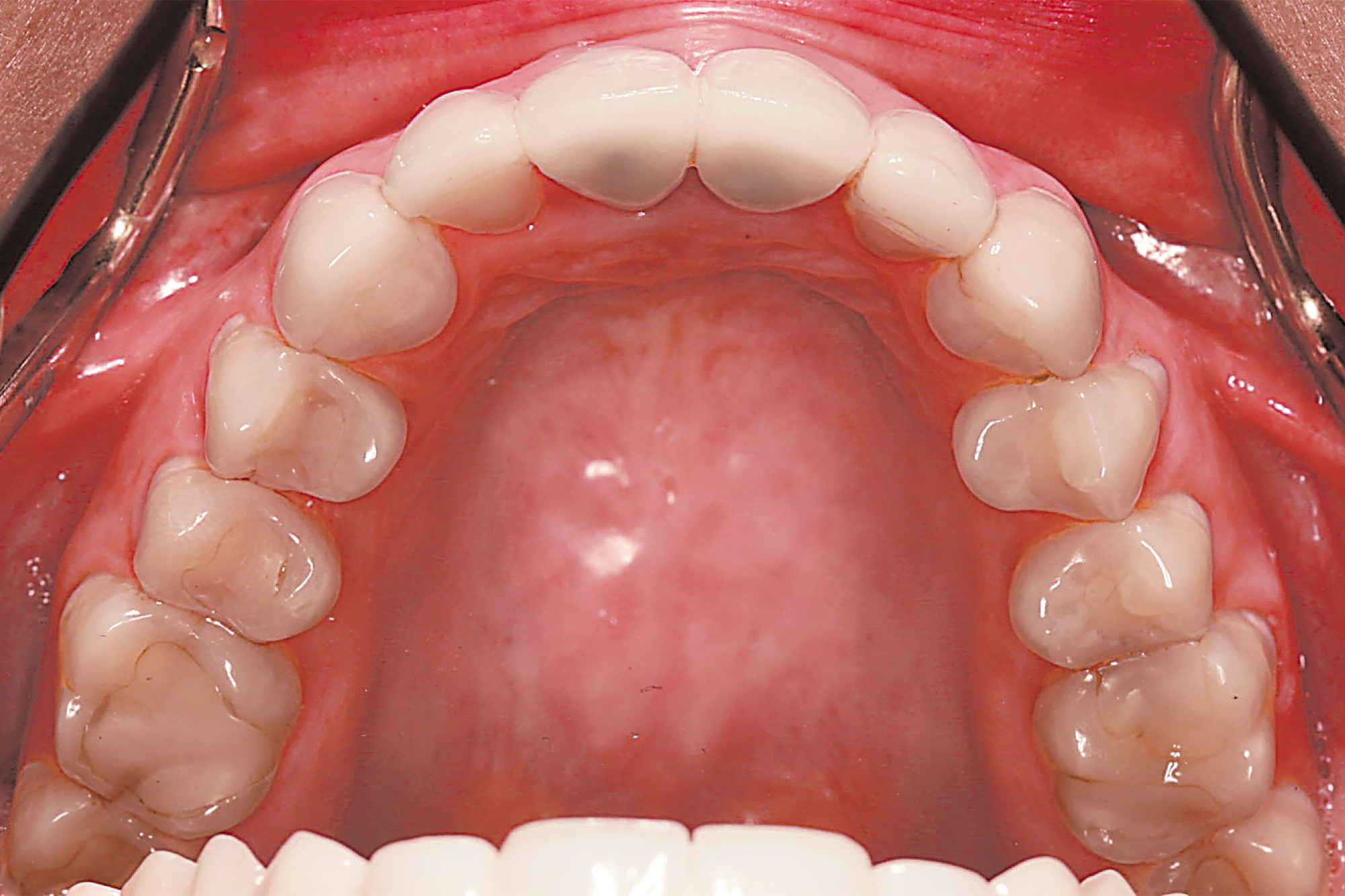
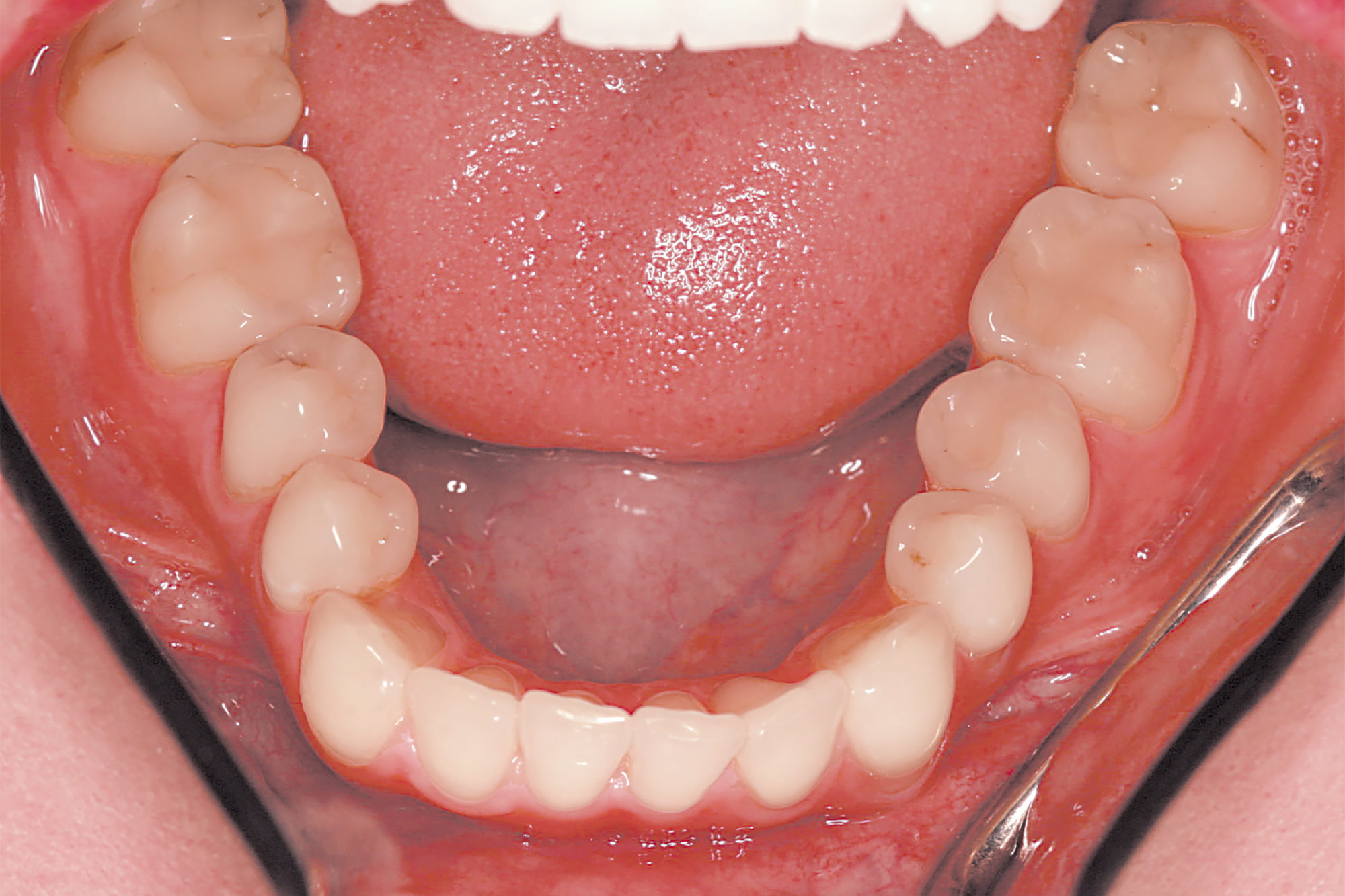
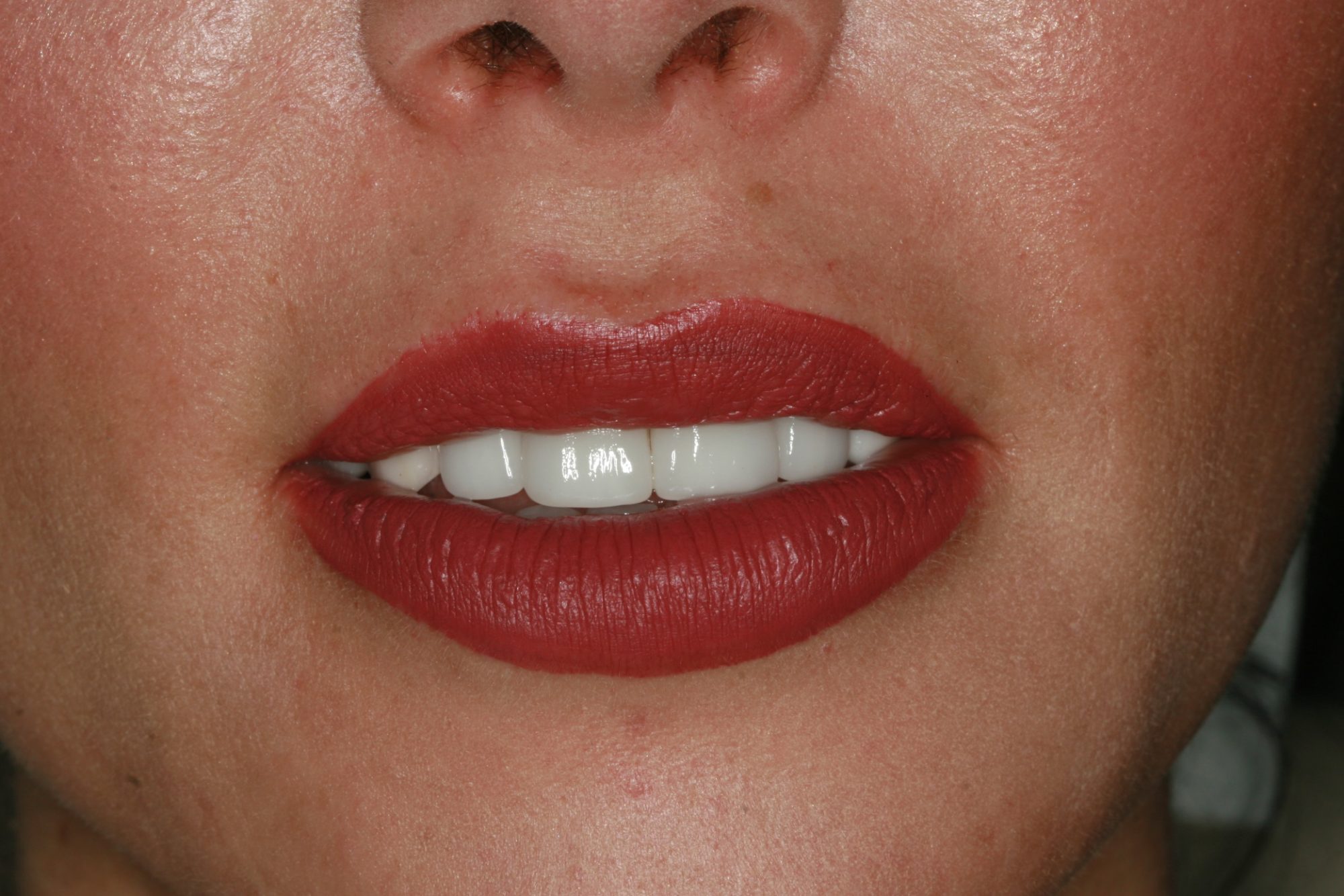
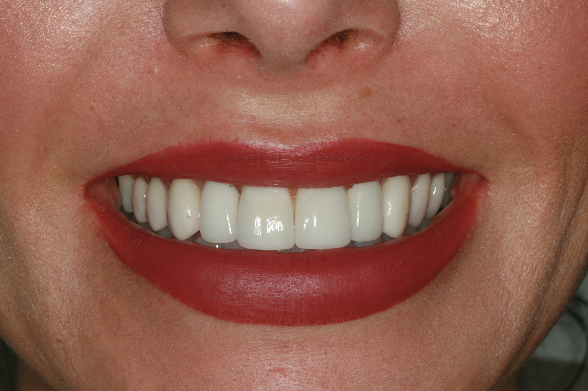
The immediate goal of treatment was to deal with the failing and infected teeth nos. 8 and 9. Once that situation was stabilized, the functional goal was to provide a long-term solution for the replacement of missing teeth nos. 8 and 9 and correct the malocclusion. The patient’s esthetic goals were to make the anterior teeth wider and longer, create a wider smile, correct the malpositioned tooth no. 27, and replace the unesthetic Class V restorations.

Phase I: Tooth Extraction and Provisionalization

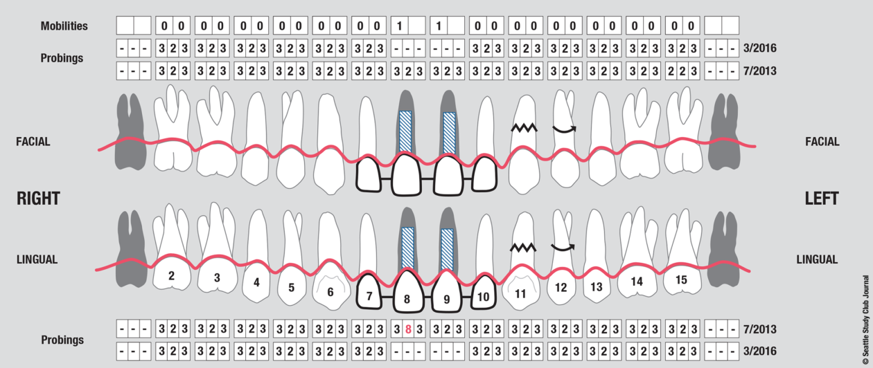
Teeth nos. 8 and 9 were extracted, the sockets were preserved with rhBMP-2 and cancellous allograft, and a provisional from teeth nos. 7 to 10—with nos. 8 and 9 functioning as ovate pontics—was delivered.

Phase II: Surgery

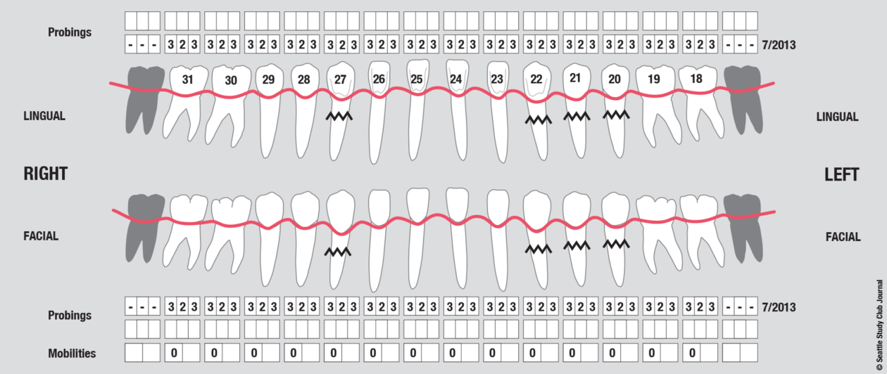
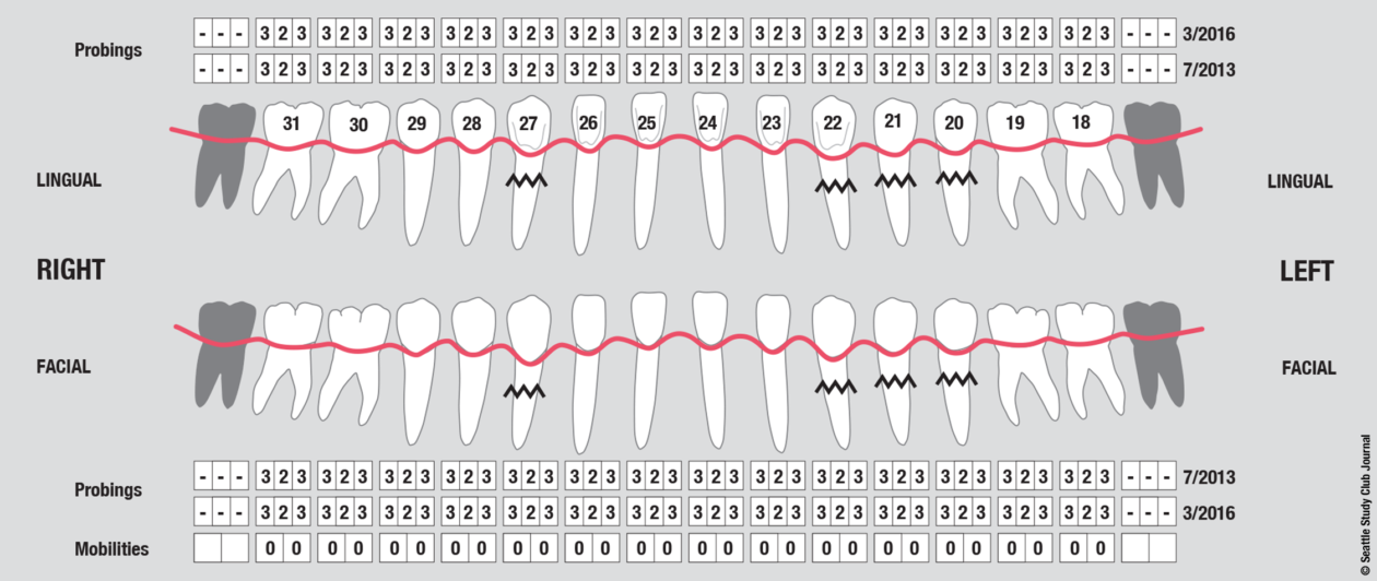
Surgically facilitated orthodontic therapy (SFOT) was initiated with corticotomies and decortication of the maxilla and mandible with the goal of expanding the arch forms and optimizing the transverse, A-P, and maxillomandibular relationships. SFOT was performed using mineralized freeze-dried bone allograft (FDBA) and Bio-Oss (Geistlich) combined with platelet-rich fibrin. Interpositional connective tissue grafting comprising autogenous tissue and Emdogain (Straumann) was undertaken for mucogingival augmentation in the areas of teeth nos. 21, 22, and 27. Horizontal and vertical tissue engineering via rhBMP-2 and cancellous allograft was completed at the sites of teeth nos. 8 and 9 for edentulous ridge augmentation.

Phase III: Implant Placement

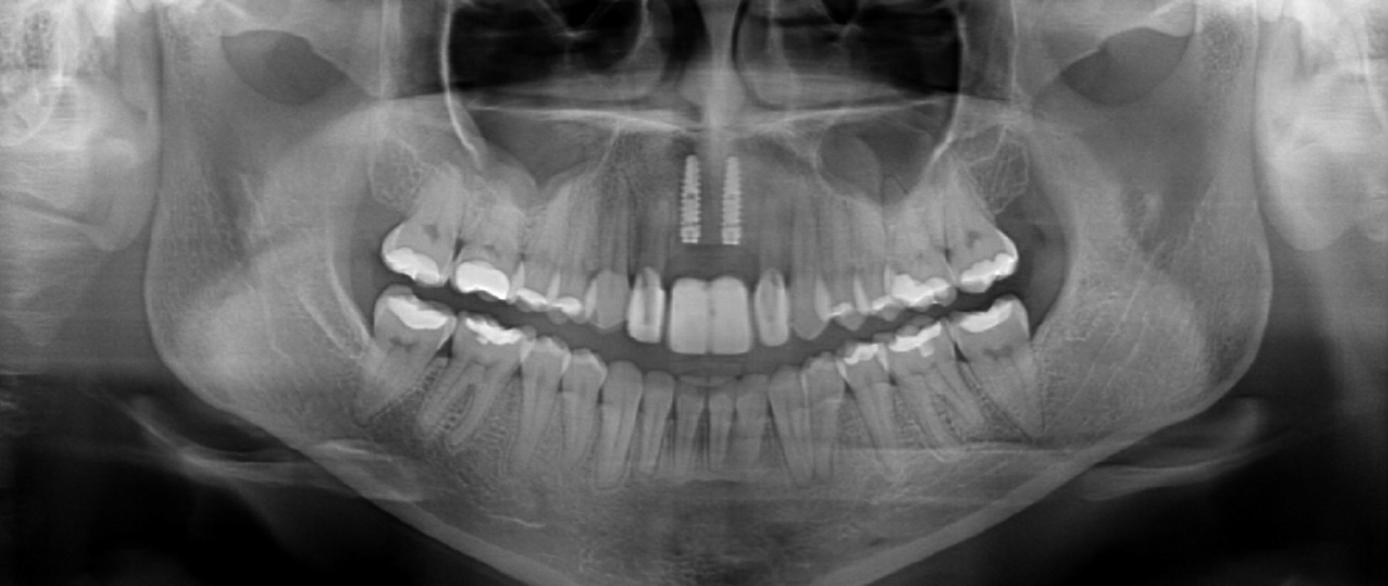
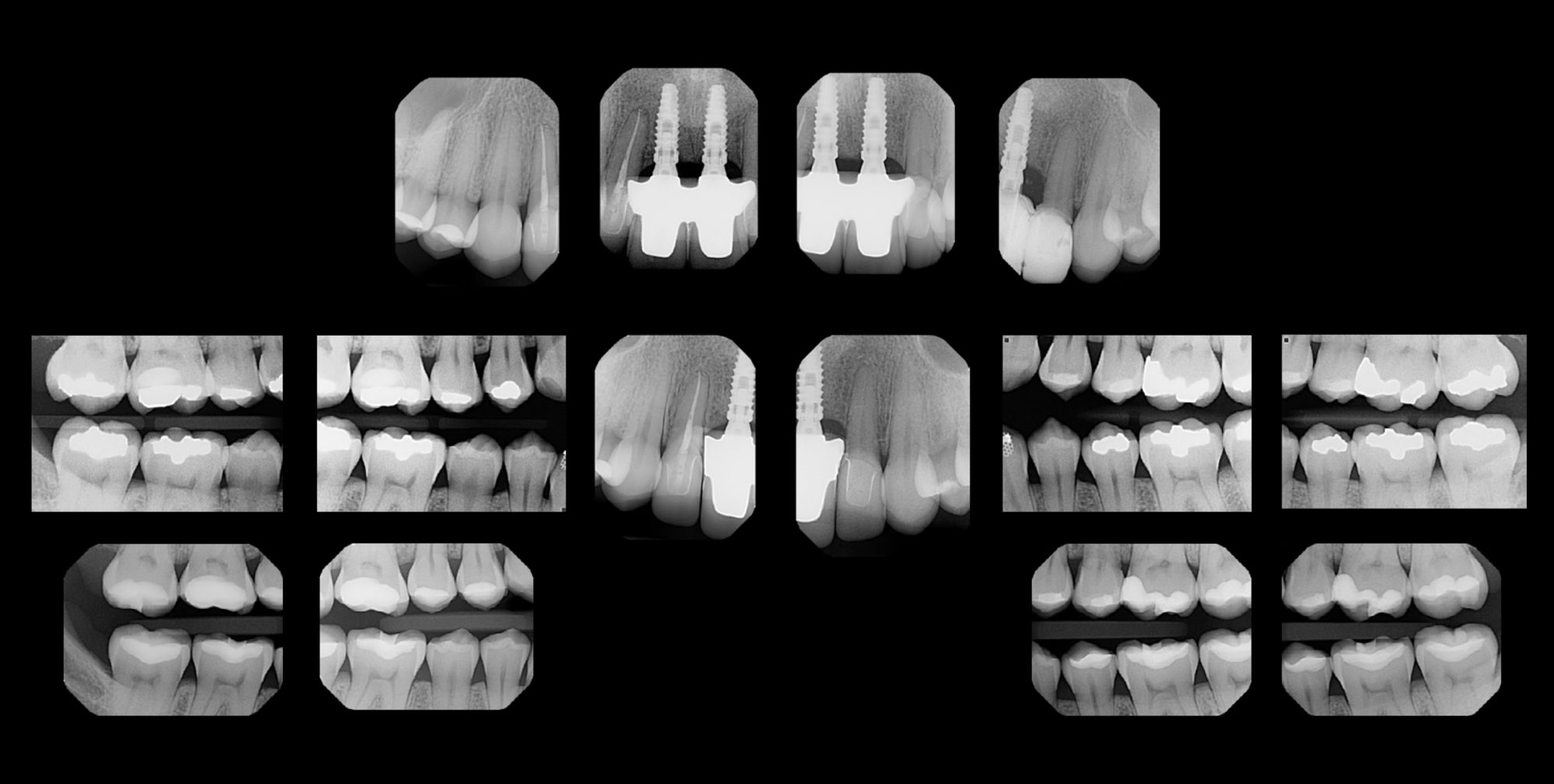
Eight months after completion of the SFOT, implants were placed (guided) at the nos. 8 and 9 sites via CBCT static guides. Contour bone grafting was performed using small-particle xenograft (Bio-Oss) + barrier membrane (Bio-Gide, Geistlich) for optimal peri-implant bone volume in combination with autogenous connective tissue for soft tissue volume. Hard and soft tissue grafting at the time of implant placement was performed to optimize the pink esthetic volume and position.

Phase IV: Placement of Healing Abutments

Four months later, temporary healing abutments were placed as “tent poles” on the nos. 8 and 9 implants and covered with an additional connective tissue graft.

Phase V: Implant Uncovering

Three months later, the nos. 8 and 9 implants were uncovered, and aberrant frenum tissue was removed.

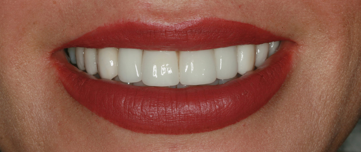
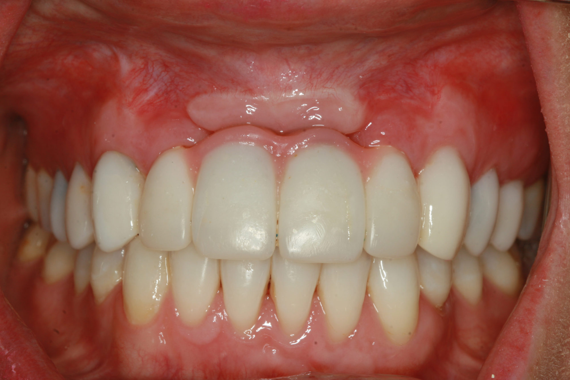
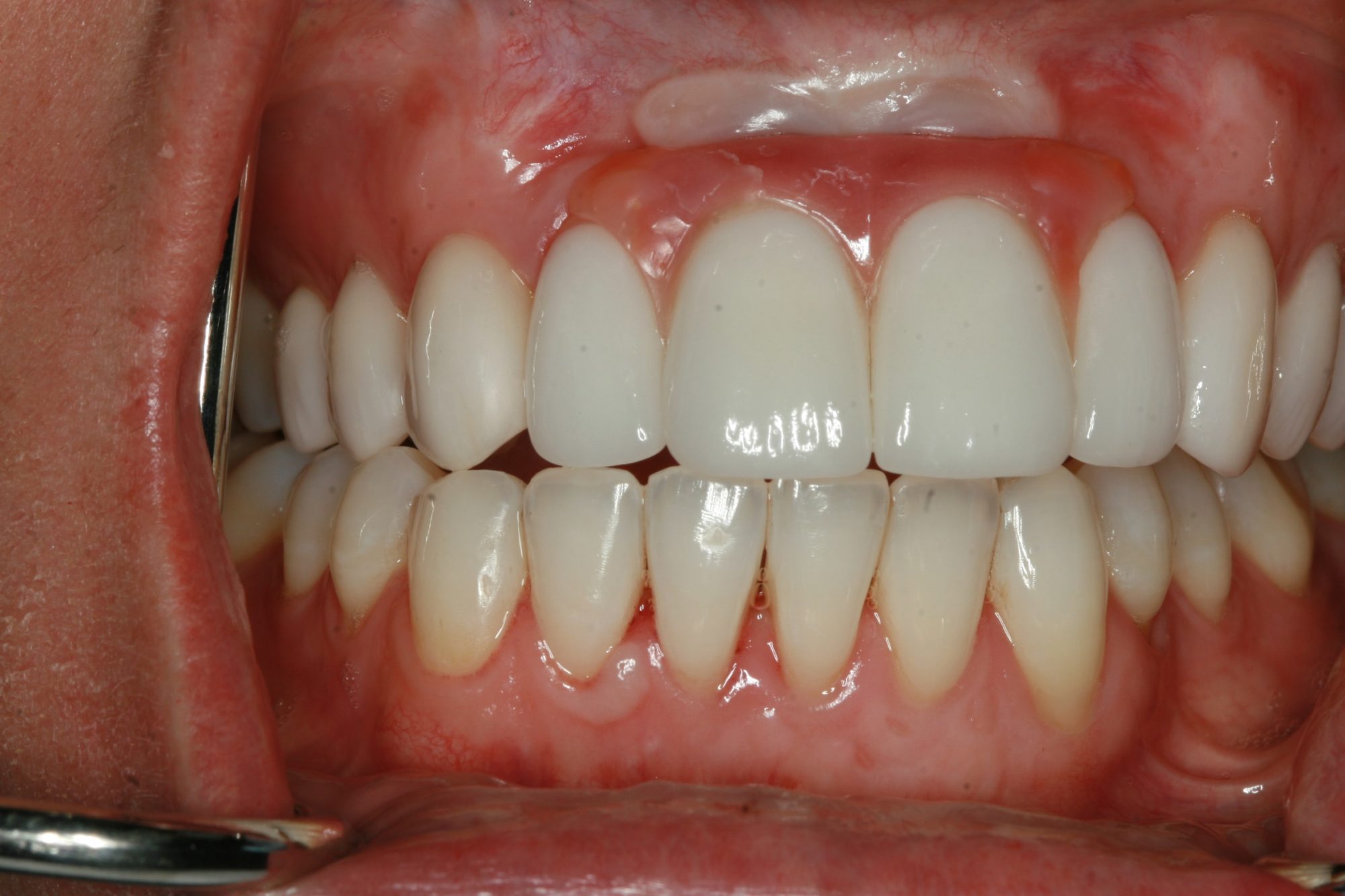
Phase VI: Implant Provisionalization (2015)

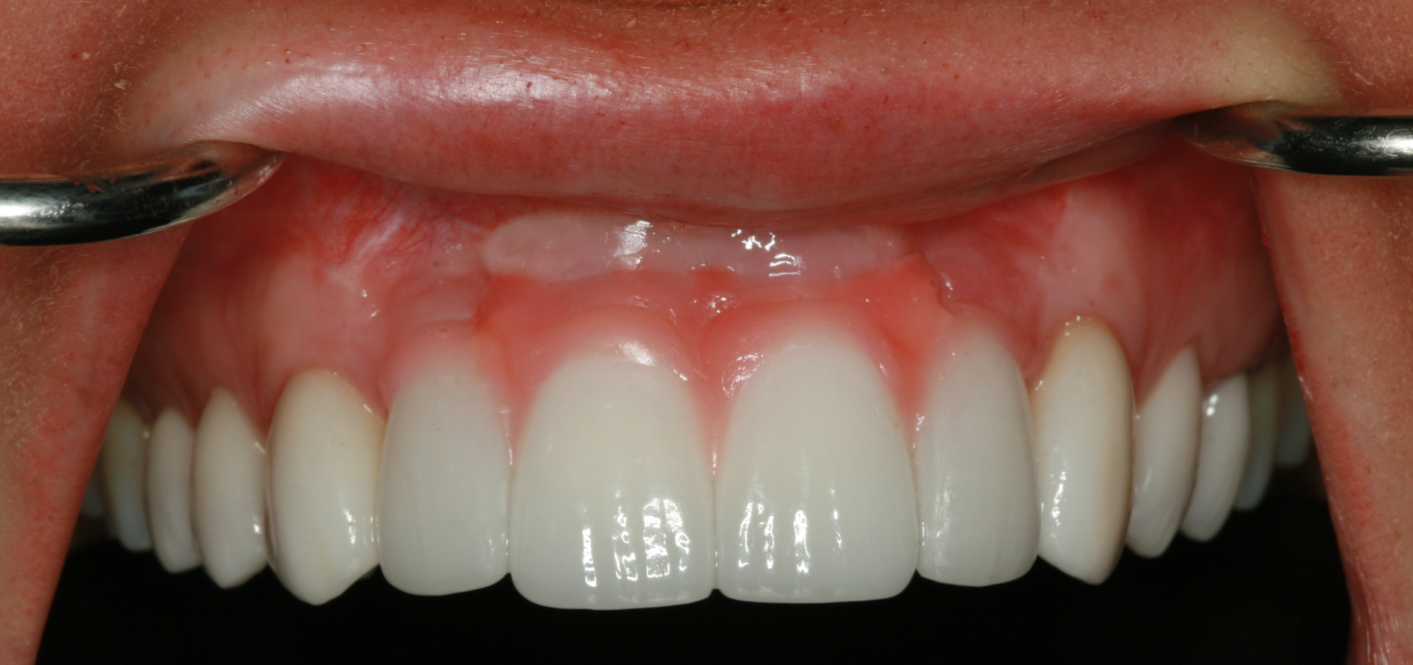
Following healing from implant uncovery, the provisionalization phase of the case ensued. This required several appointments for seating provisionals and working with contours to groom the soft tissues. The goal was to complete the case with conventional prosthetics, but despite all efforts the final bone topography did not support ideal tissue positions.

Provisional restorations in repose.
Provisional restorations light smile.
Best provisional without pink.
Pink provisional.

Phase VII: Definitive Restorations (2016)

Definitive restorations were delivered with ceramic crowns on nos. 7 and 10 and a splinted zirconia framework that had pink porcelain and pink composite layered on the facial aspects of the central incisors. This also established the midline papilla. In addition, there were wings that overlaid the facial of the lateral incisors to create the balanced papilla heights necessary to meet the patient’s esthetic demands. The splinted zirconia prosthesis with pink porcelain and composite was used to overcome the shortcoming in gaining optimal hard and soft tissue volume dimensions in the anterior maxilla.

Phase VIII: Maintenance (2016–)

The patient is on a 3- to 4-month recall program at which annual radiographs are taken of the implants to ensure maintenance of the peri-implant bone anatomy. Her home care is quite good, and she is able to maintain favorable hygiene in the anterior maxilla. She is able to floss under and around the pink wings on the mesial and facial aspects of the lateral incisors and maintain a healthy and plaque-free environment for the entire prosthesis.

Definitive repose.
Definitive high smile.
Definitive right working.
Definitive left working.
Definitive full face.
Definitive profile.

Commentary

This case did not turn out as originally planned, which is the primary reason we decided to present it here. Relatively complex treatment was rendered with the goal of obtaining ideal arch forms, maxillomandibular relationship, anterior-protected articulation, and especially the intrinsic proportions of the teeth (white esthetics) by optimizing the position of the pink esthetics developed through reconstructive hard and soft tissue surgery.

The objectives of employing SFOT as part of the interdisciplinary therapy were the following:

  • To put the teeth in the correct position for facial esthetics and function.
  • To transform and enhance the dentoalveolar bone phenotype in order to make goal no. 1 possible without creating iatrogenic periodontal problems.
  • To optimize anterior-protected articulation parameters.
  • To reduce the incidence of orthodontic relapse by ensuring that the teeth were completely housed in alveolar bone following orthodontic expansion.
  • To optimize airway conditions by increasing the oral cavity volume.

From a surgical perspective, our plan and sequence seemed ideal, well-crafted, and novel. However, by taking on more complex treatment, we also suffered the equally complex problems that are highlighted here:

  • During the orthodontic expansion, which was undertaken to obtain the width required for the digital smile-designed central and lateral incisors, the bone anatomy flattened and the interproximal peaks of bone supporting the papillae were compromised.
  • Healing from the maxillary SFOT and guided bone regeneration (GBR) surgery using tissue engineering progressed uneventfully. However, because of the need for primary wound closure in the anterior maxilla, the flap was significantly advanced for tension-free closure, and consequently the mucogingival junction became distorted.
  • As space for the central incisors was developed, we lost vertical bone height, further complicating our efforts to maintain the interproximal peaks of bone at the sites of nos. 7–8, 8–9, and 9–10. Ideally, a secondary GBR surgery might have been considered to gain more ideal crestal bone volume in both vertical and horizontal dimensions and, ultimately, support the desired pink esthetics.

In such complex and demanding GBR surgery, it is common to encounter mucogingival problems. Usually, these require soft tissue augmentation by means of a strip graft, or apical repositioning of the flap. In this case, we decided to correct the volume deficiency with a second attempt at interpositional connective tissue grafting because the implants appeared to have favorable circumferential bone volume following the contour bone grafting that was performed simultaneous with connective tissue grafting and guided implant placement. At the implant augmentation surgery, some shrinkage in bone or soft tissue volume was expected. The problem in this case is that despite multiple attempts to gain the ideal volume, we came up short. After orthodontia and several surgeries, the risk of depleting the emotional currency of the patient threatened to become a management issue (a subject that is not often discussed), especially considering that significant prosthodontic procedures were required before case completion. In retrospect, it may have been more ideal to either (1) limit the expansion that occurred and use the same tooth proportions, which would have necessitated smaller-diameter implants, or (2) consider a secondary GBR procedure to better support the soft tissue parameters. In fact, forced eruption of tooth nos. 8 and 9 could have been considered to gain some soft tissue volume before extractions were performed. Originally, we had also discussed the option of resecting the anterior maxilla to allow for a planned pink porcelain hybrid design, but we decided against it because we believed that the regeneration needs could be met.

When it came to uncovering the implants, we were faced with a continuing pink esthetic volume deficiency and an aberrant frenum with unfavorable muscle position. We decided to uncover the implants and place an autogenous soft tissue graft to prevent the frenum from re-attaching as well as improve the surface characteristics of the anterior gingiva. This created a less-than-ideal outcome and did not solve the volume deficiency from which we needed to recover. The prosthesis design we ended up with also will require more demanding maintenance and patient hygiene and will increase the risk of peri-implant problems if the patient is unable to maintain an optimal plaque control regimen. Multiple appointments were made in the provisionalization phase to attempt to gain optimal tooth form and proportion using natural tooth form before we decided on the addition of pink porcelain to compensate for peri-implant deficiencies. Multiple appointments and videos of the patient speaking were also needed to optimally position the incisal edges in the smile design of this case. The videos helped confirm the decision to use pink porcelain and composite for optimal pink contours (see video on page 13).

In the final analysis, the patient ended up with a beautiful result, although not what was originally planned or intended. Our goal in presenting this case is to show the everyday realities of complex interdisciplinary treatment planning and highlight how a team manages adversity when nature and biology undermine our best planning efforts.

Assessment of the esthetic and phonetic contours of the provisional restorations.

George Mandelaris, DDS, MS, is in private practice in Chicago, Park Ridge, and Oakbrook Terrace, Illinois. He limits his practice to periodontology, dental implant surgery, bone reconstruction, and tissue engineering surgery.

Nader Sharifi, DDS, MS, currently maintains a full-time private practice of prosthodontics in Chicago’s downtown loop.