Case 102: In a World Where Implant Dentistry Dominates, Sometimes Traditional Periodontal Treatment Is Still Best

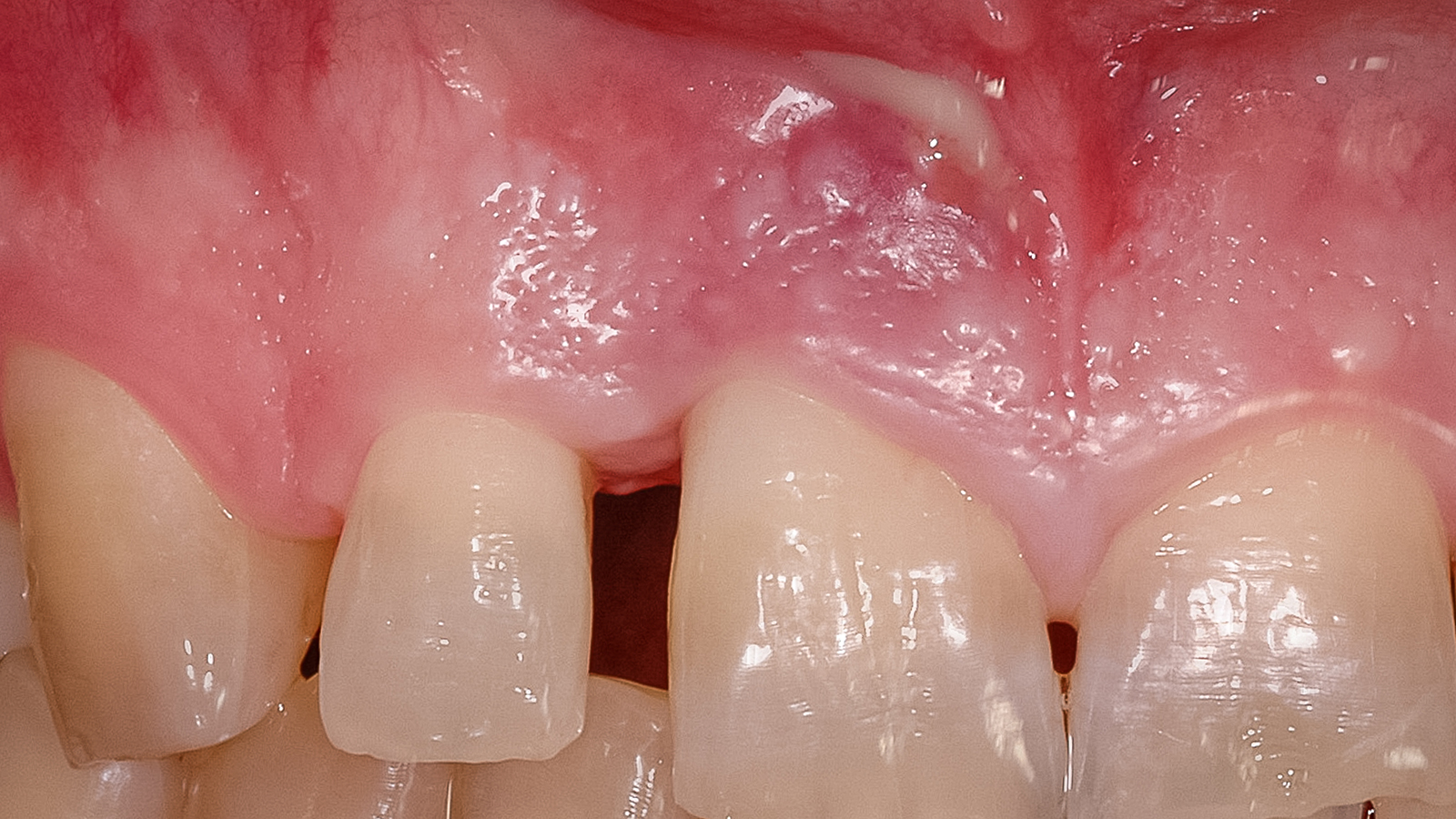
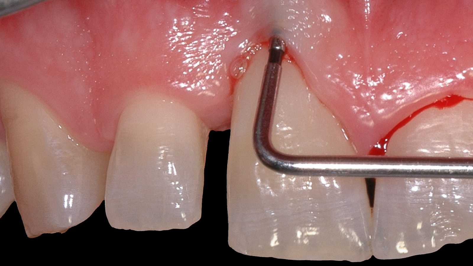
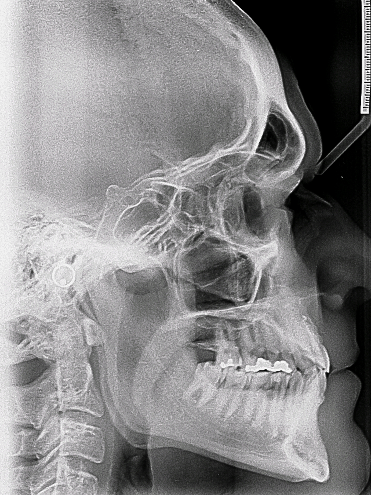
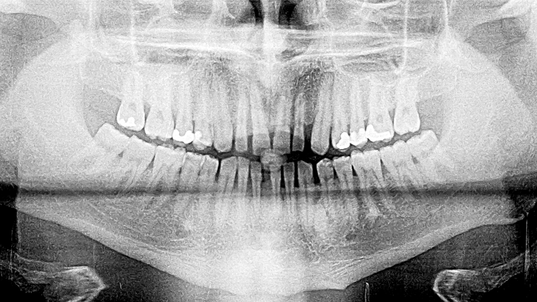
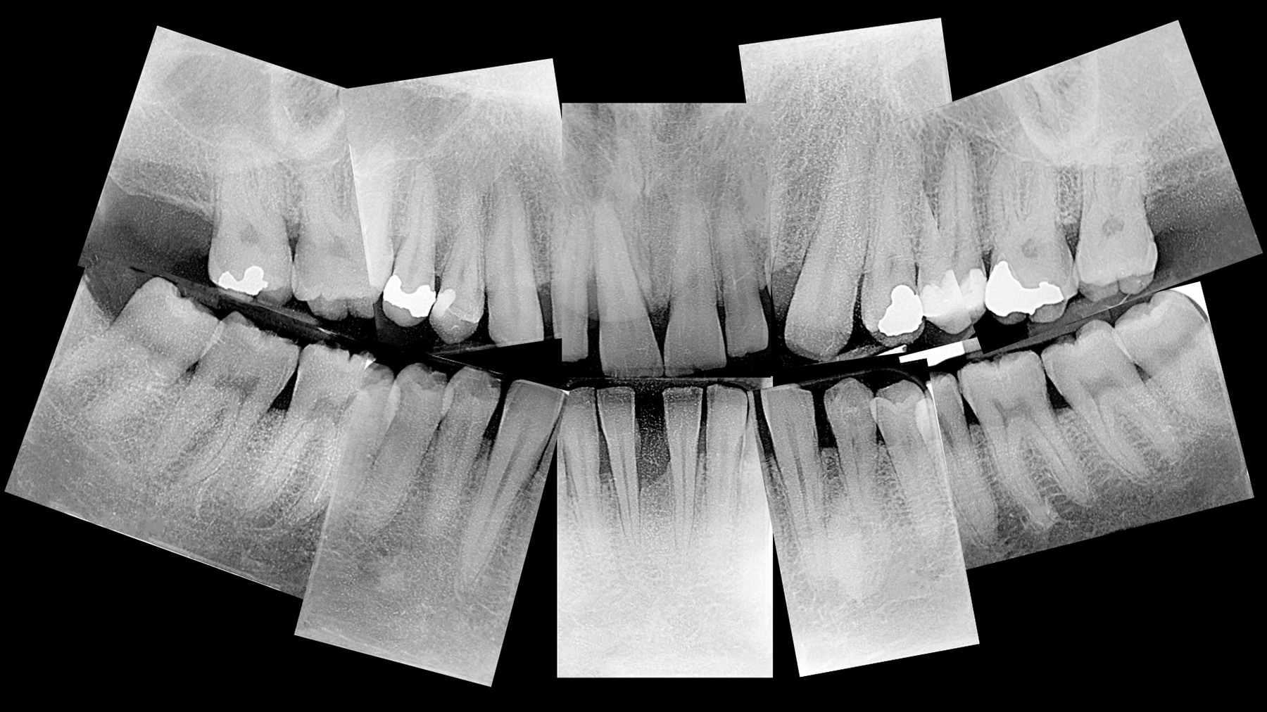
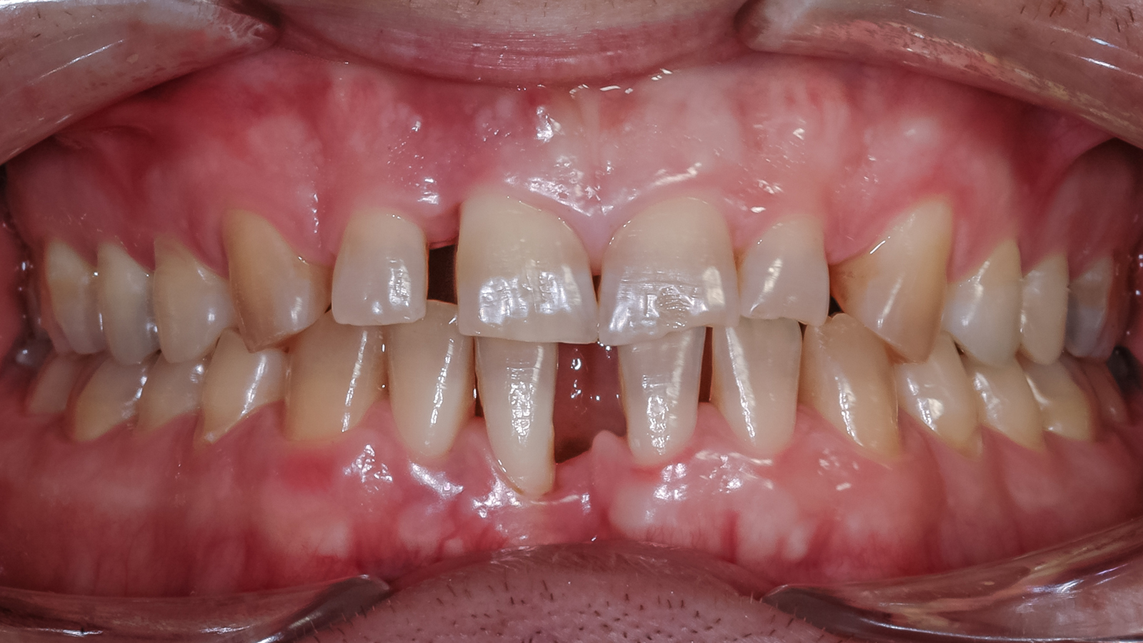
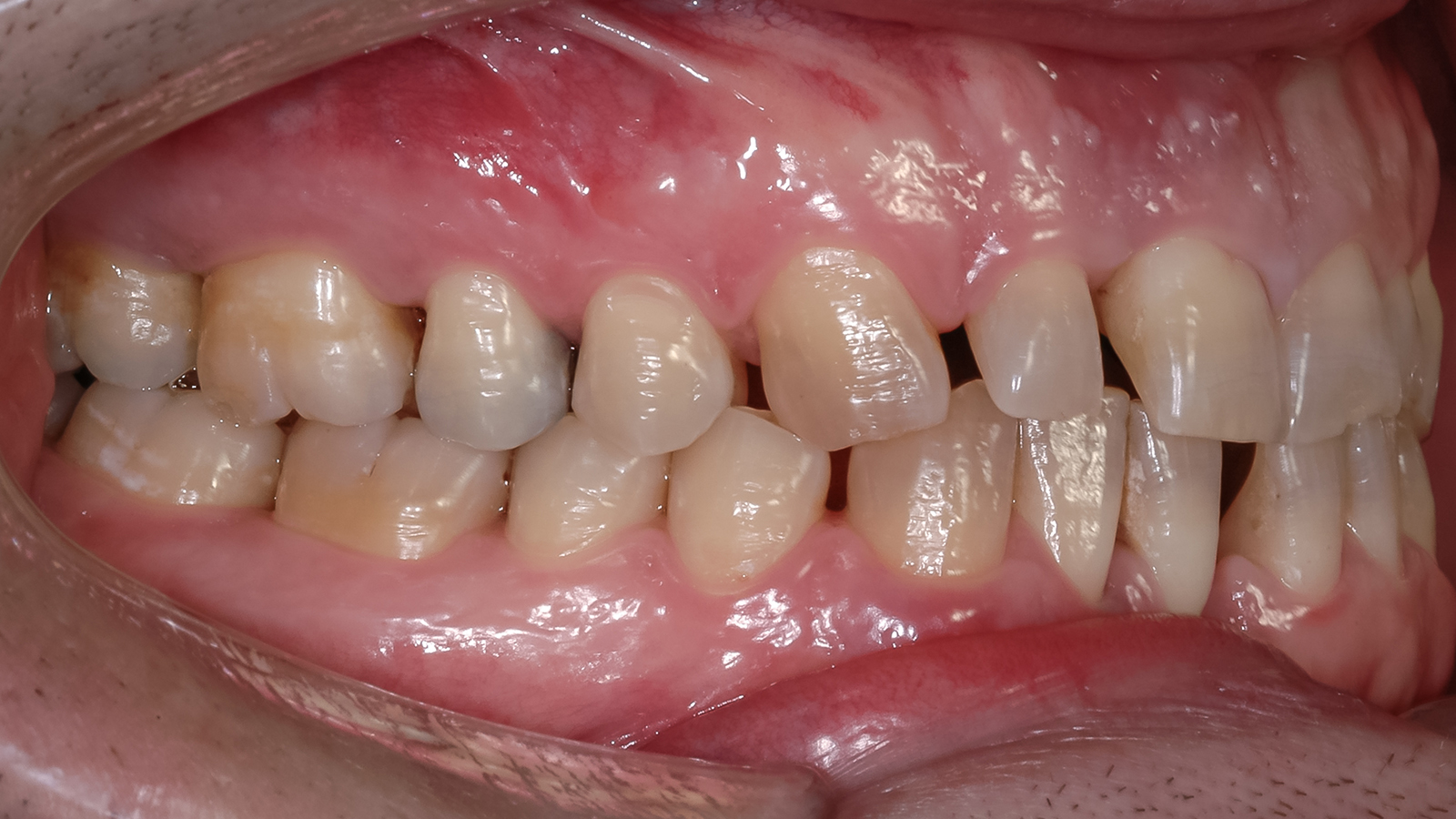
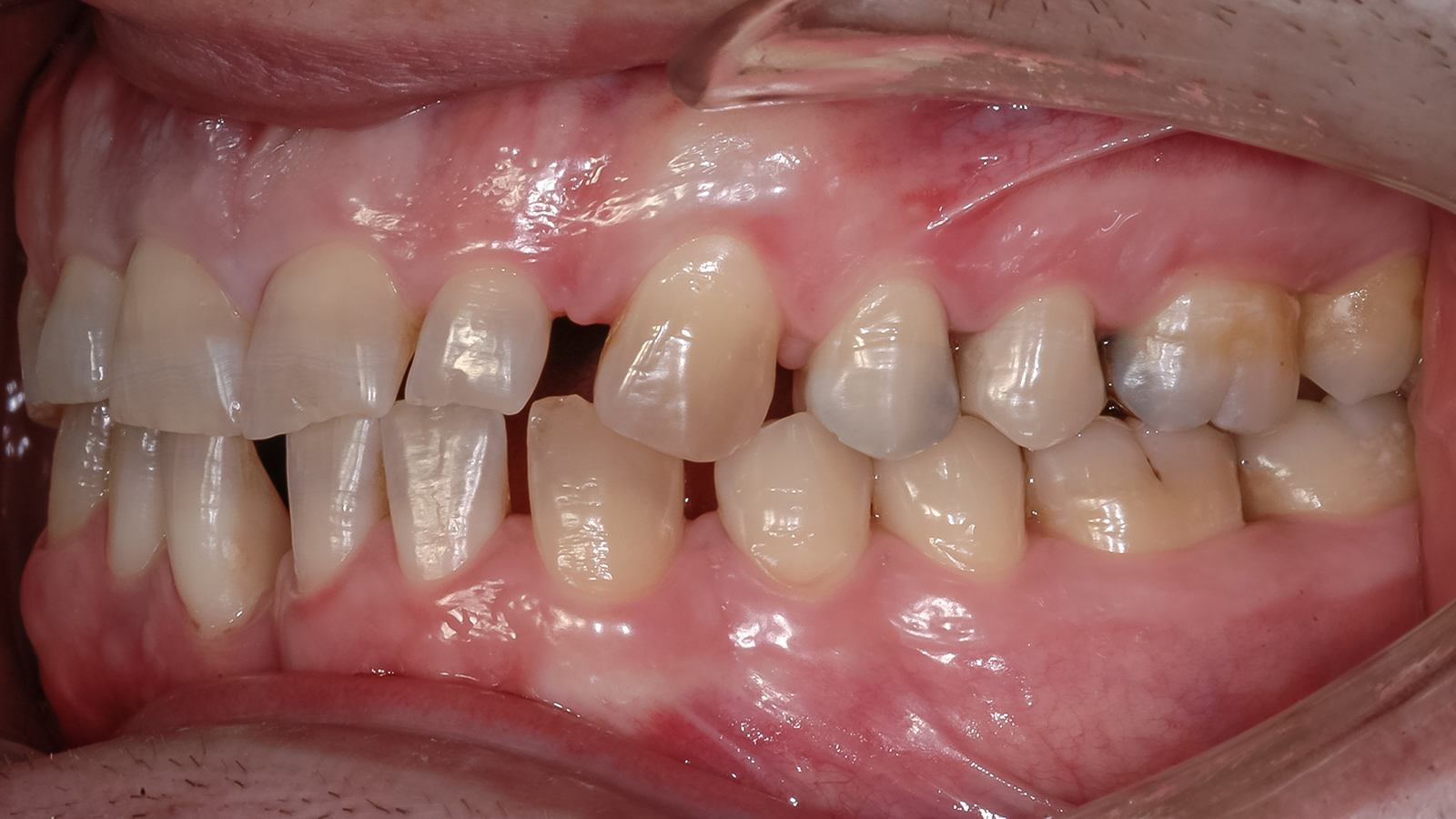
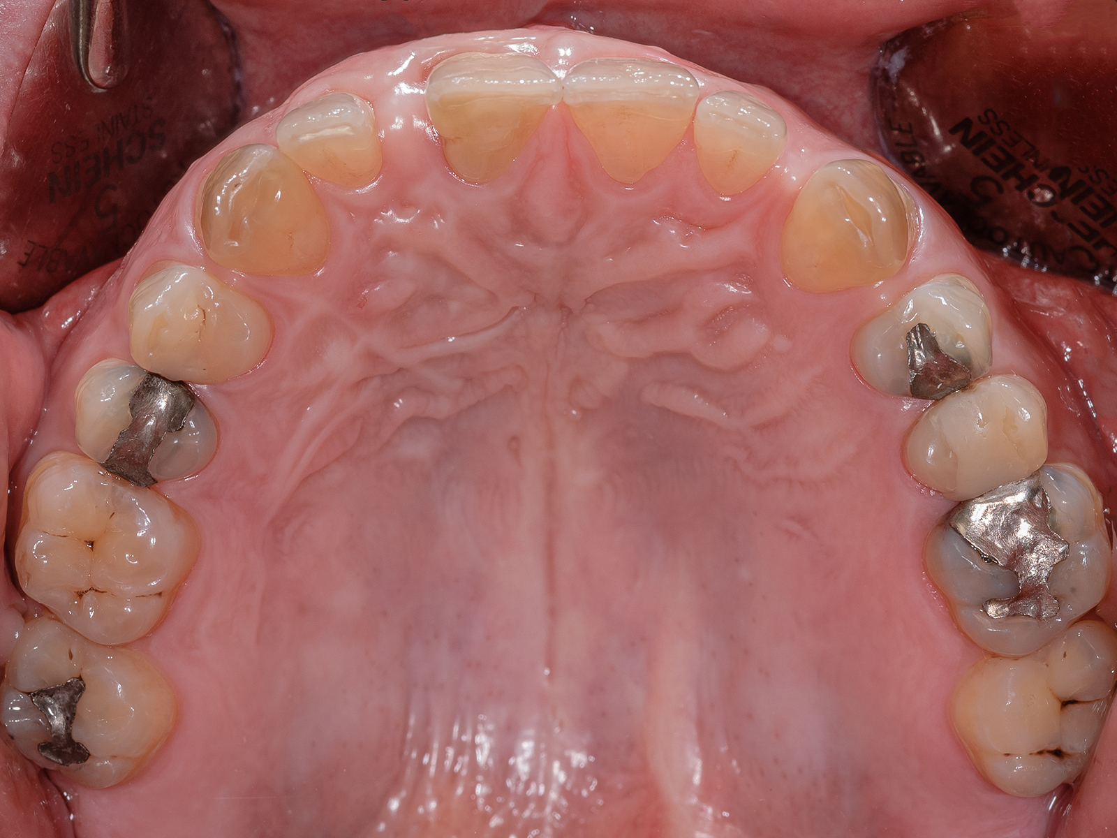
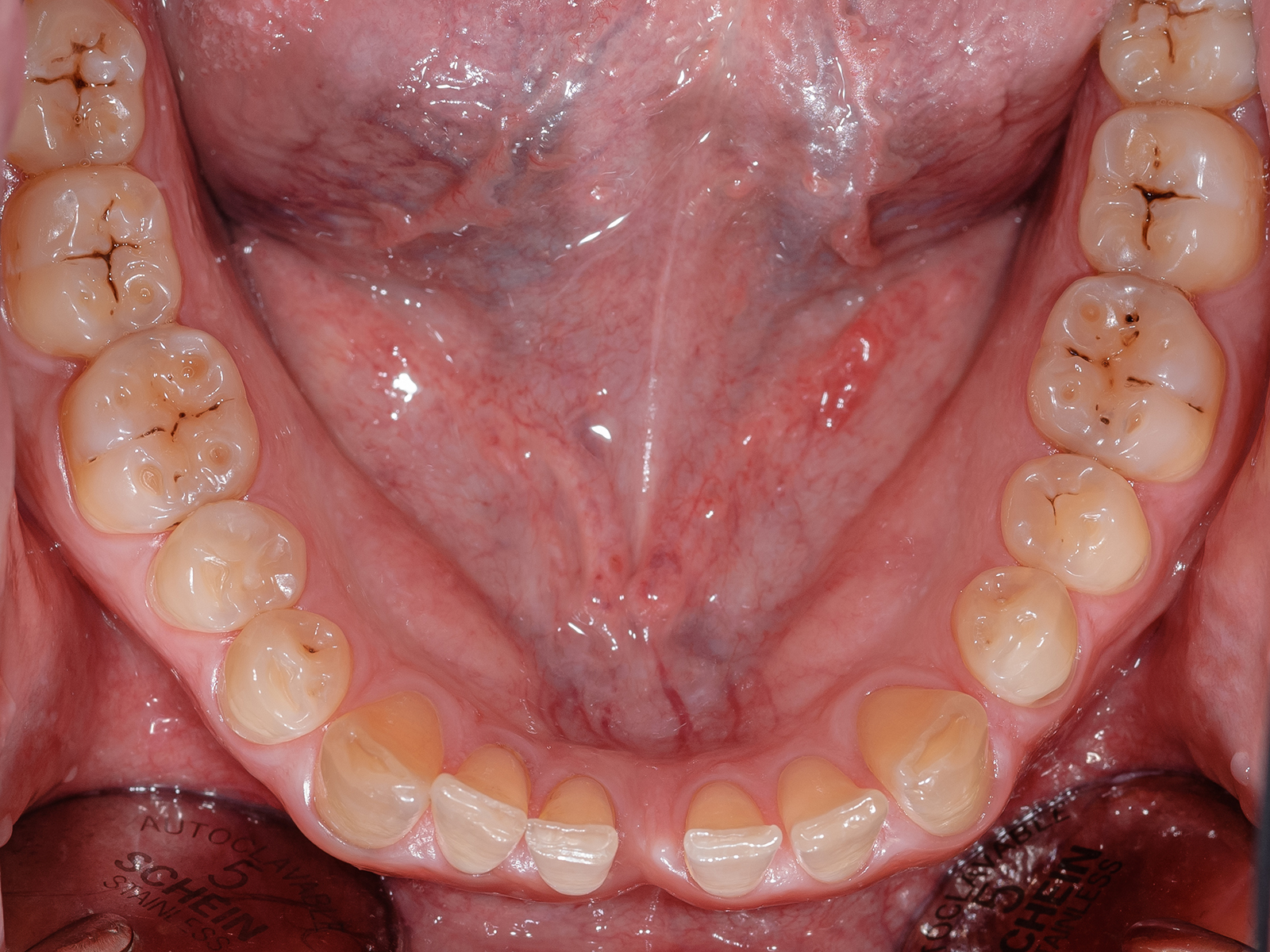
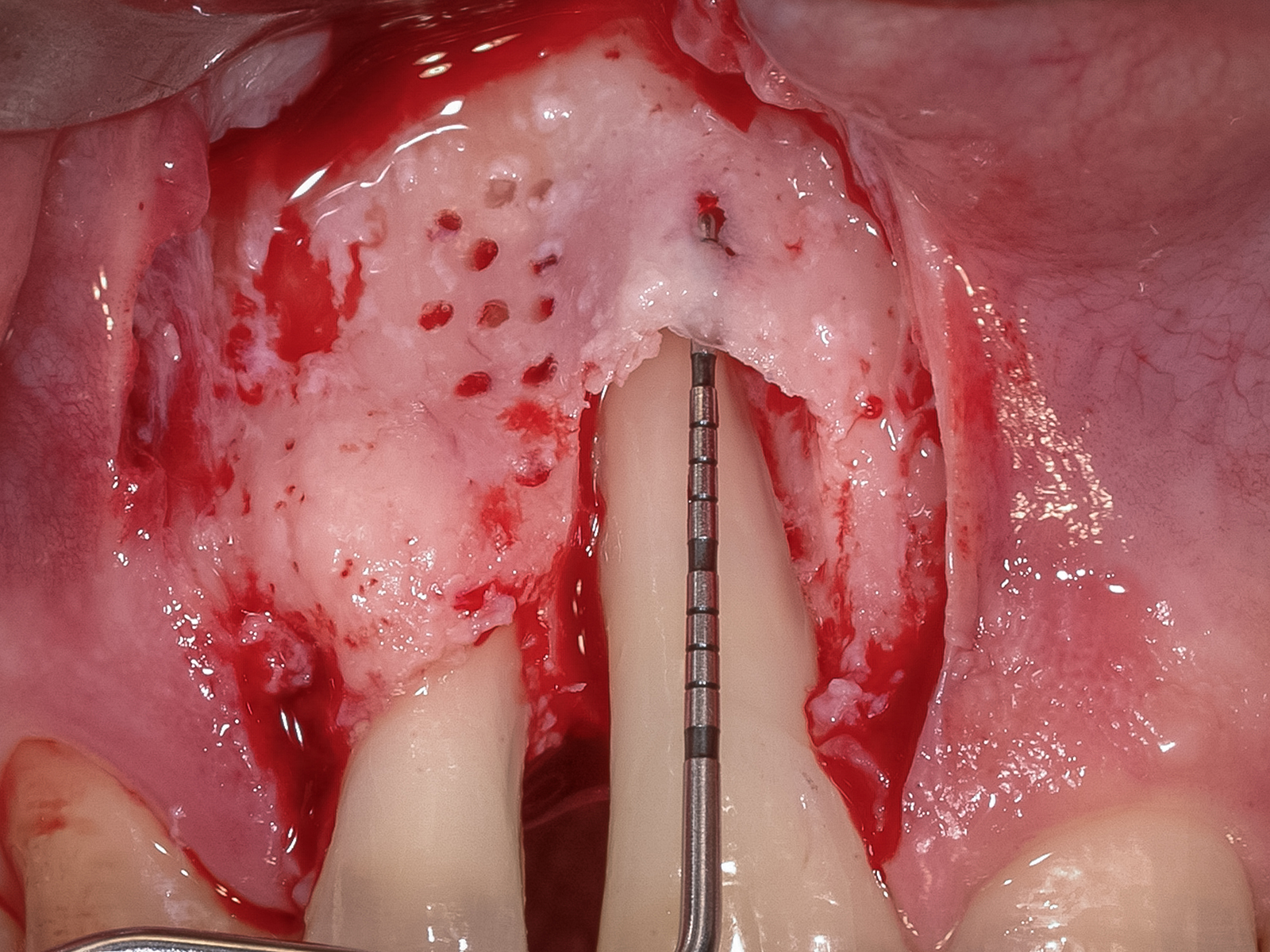
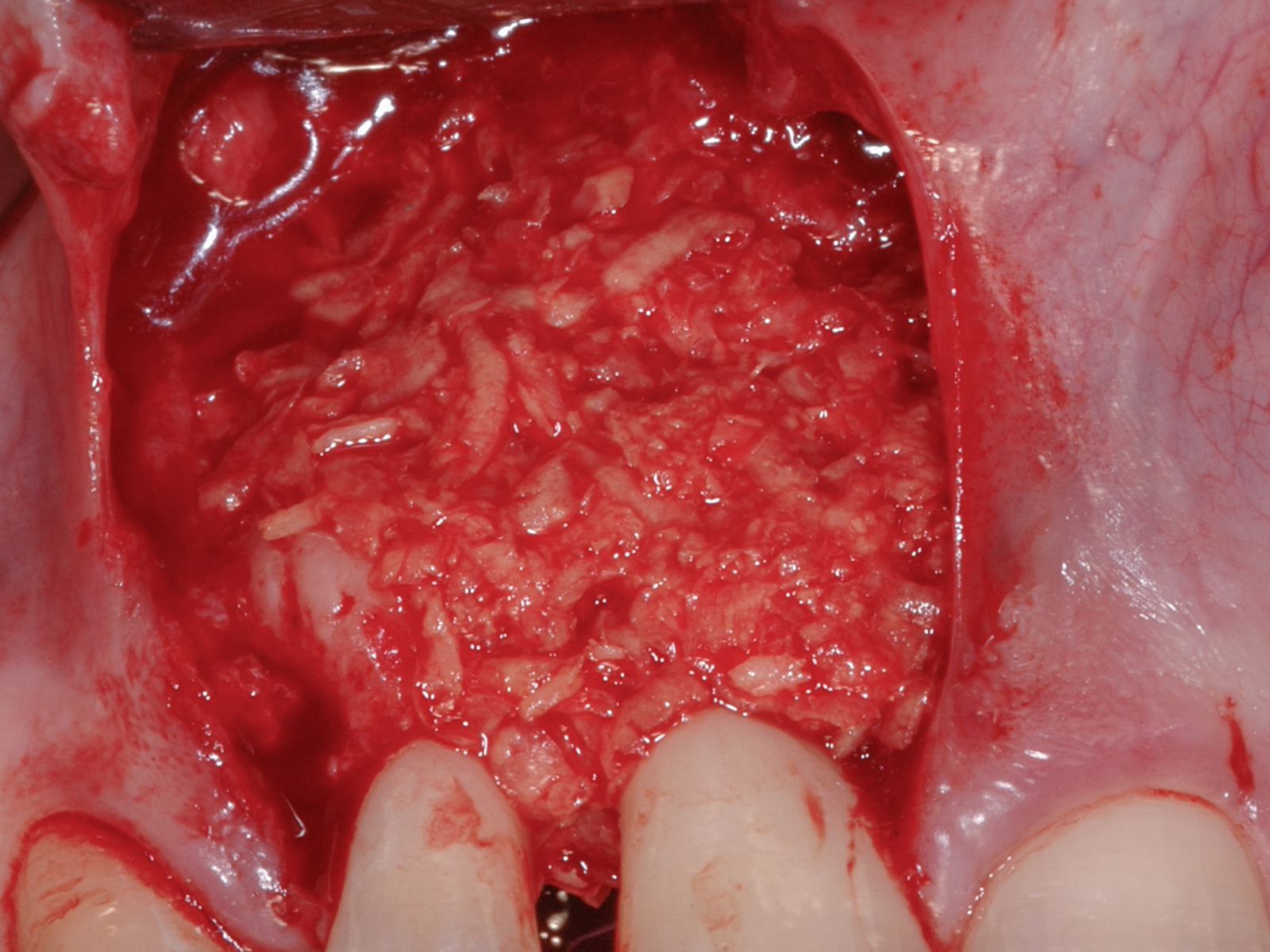
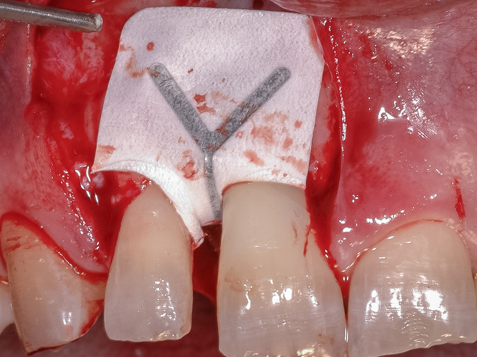
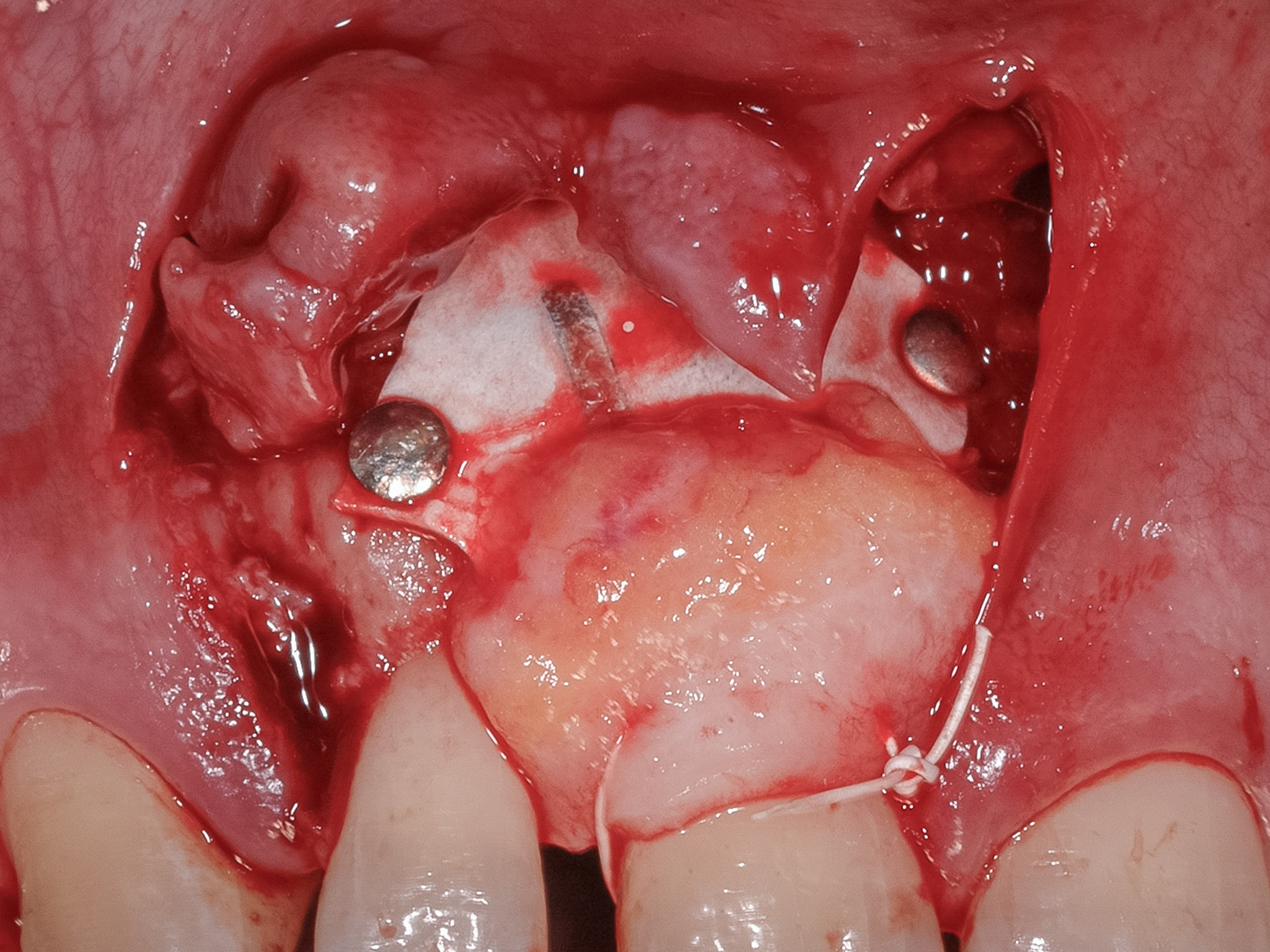
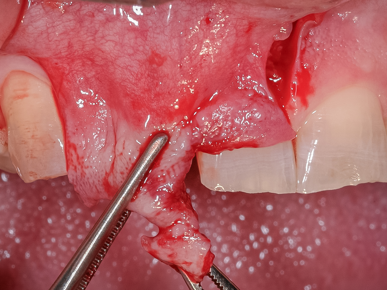
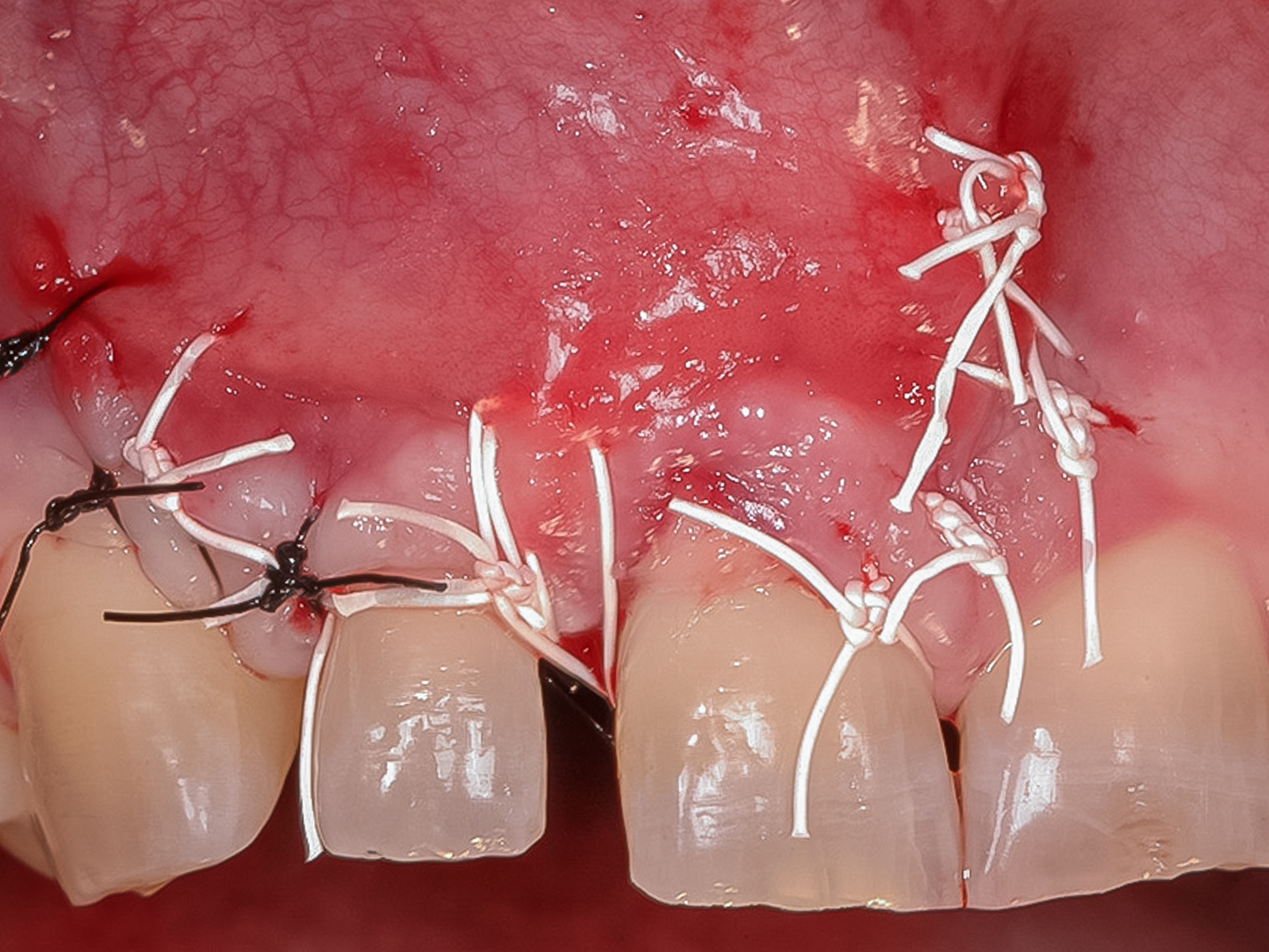
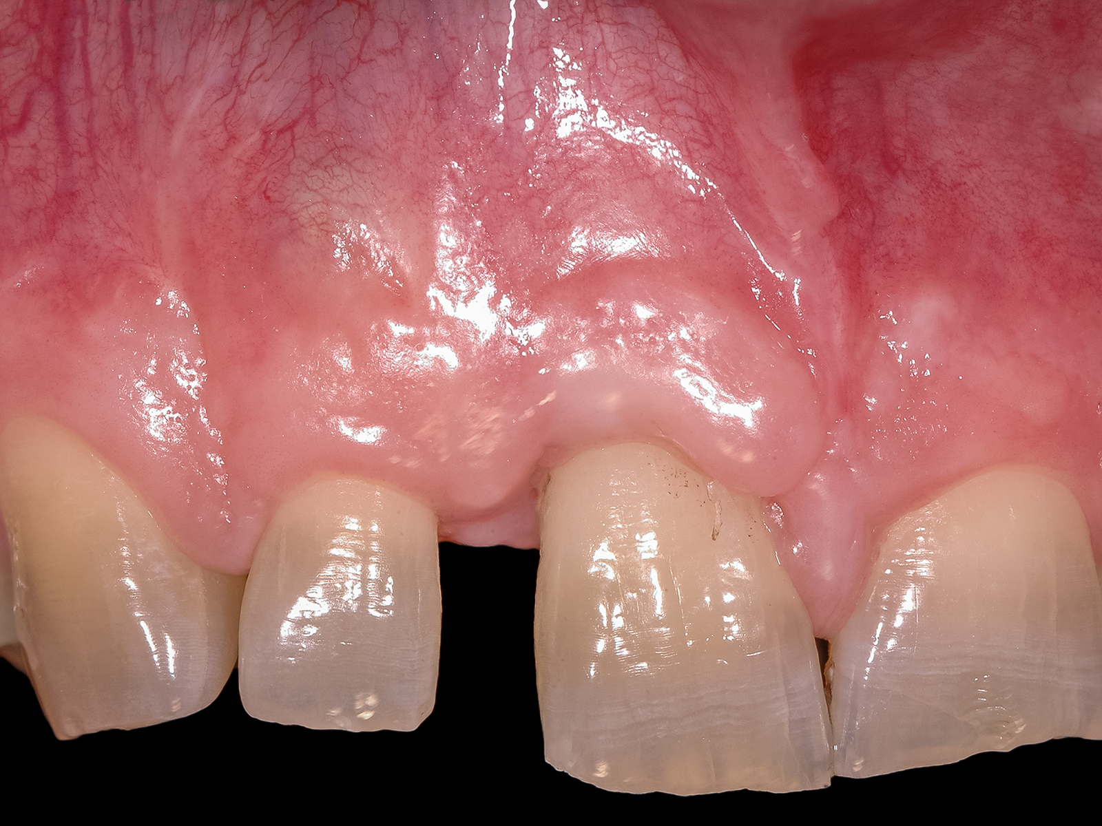
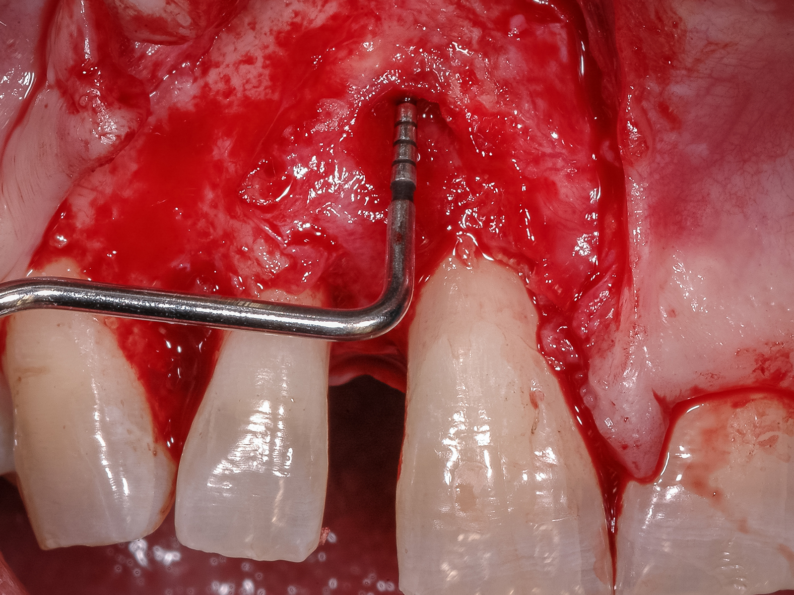
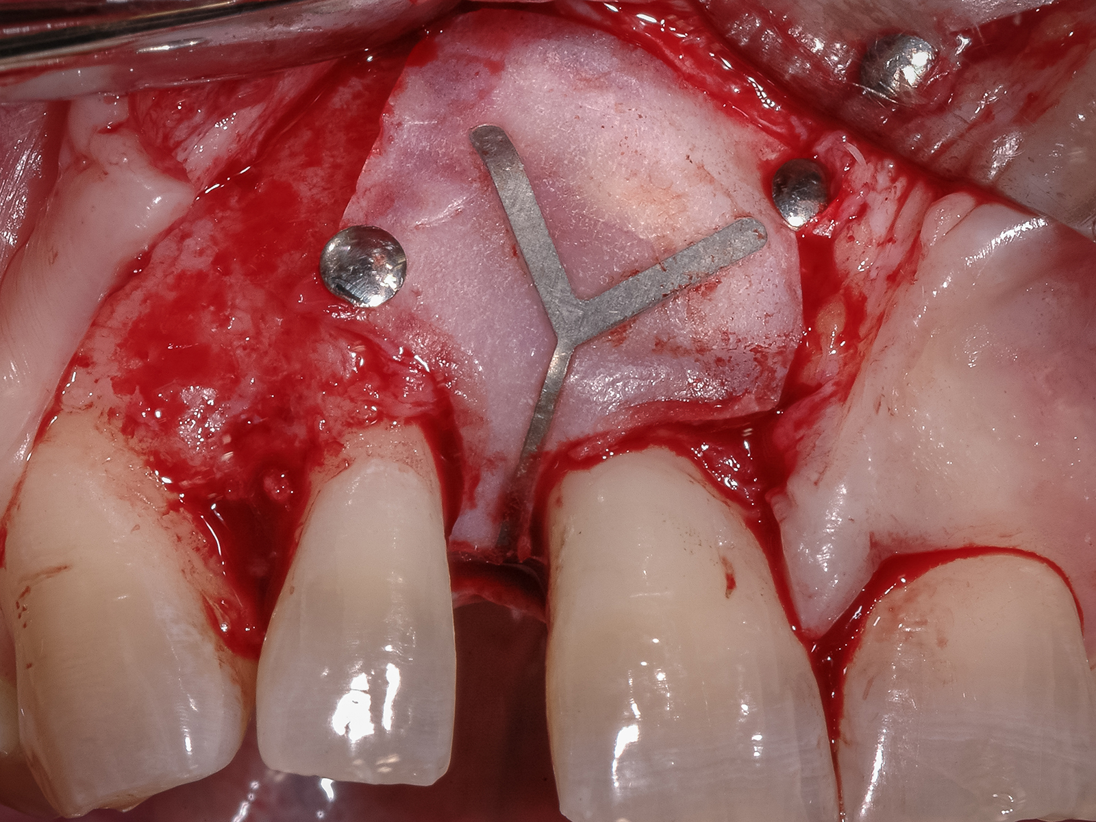
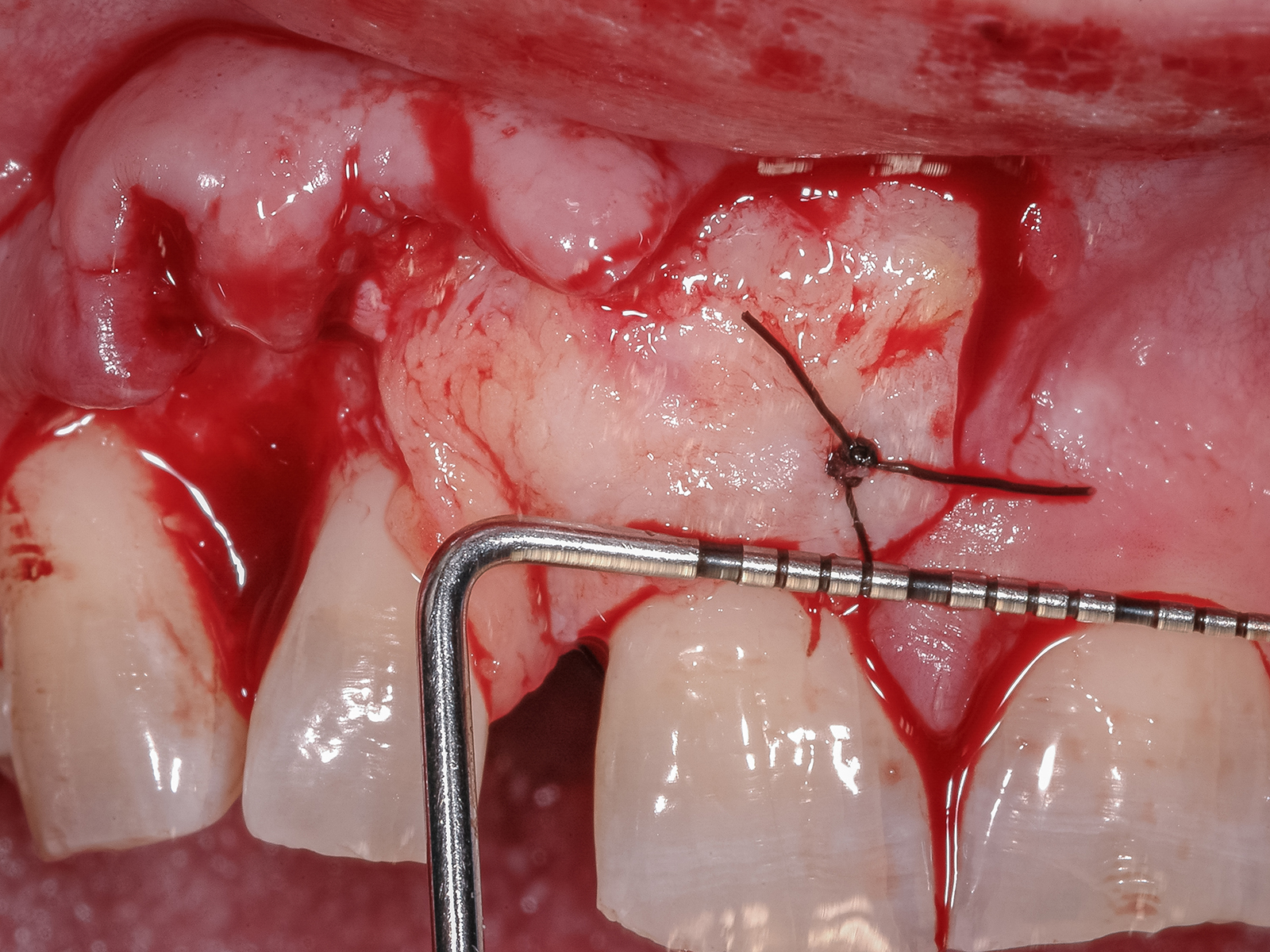
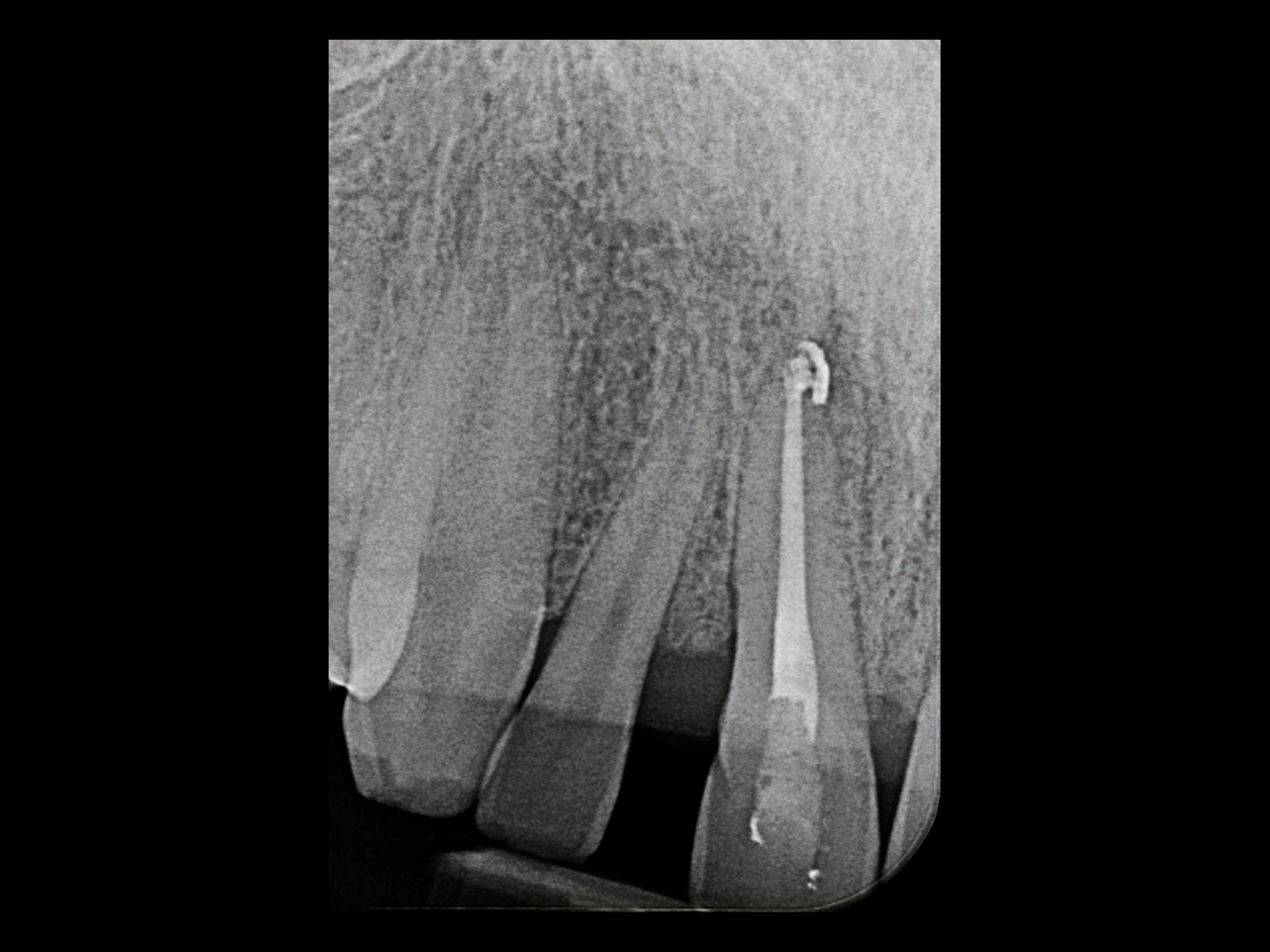
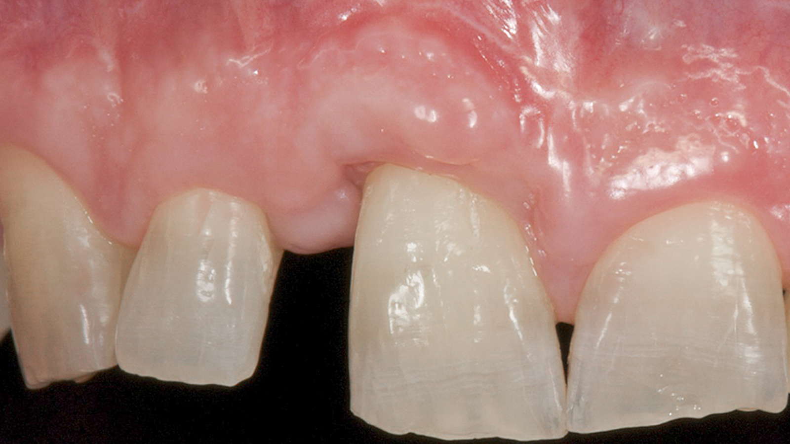
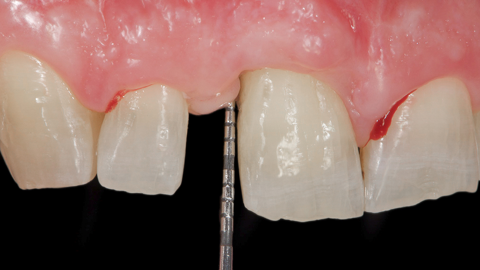
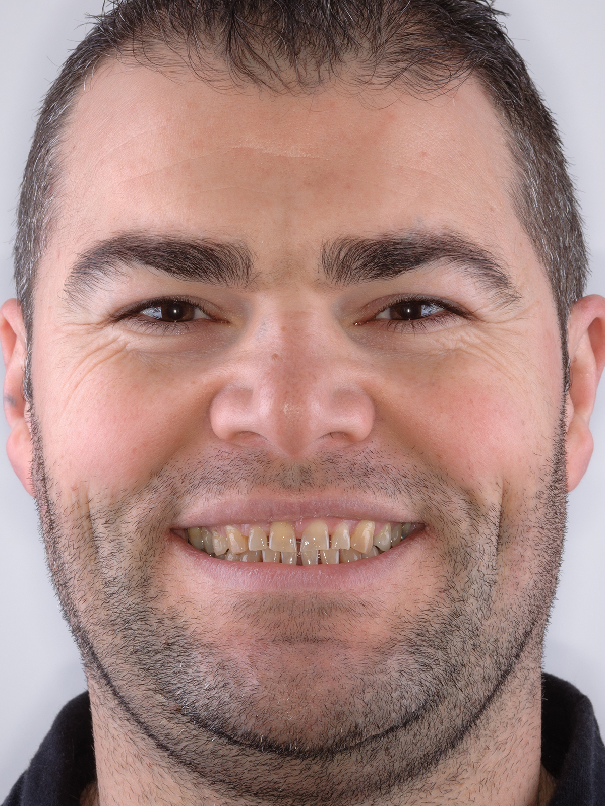
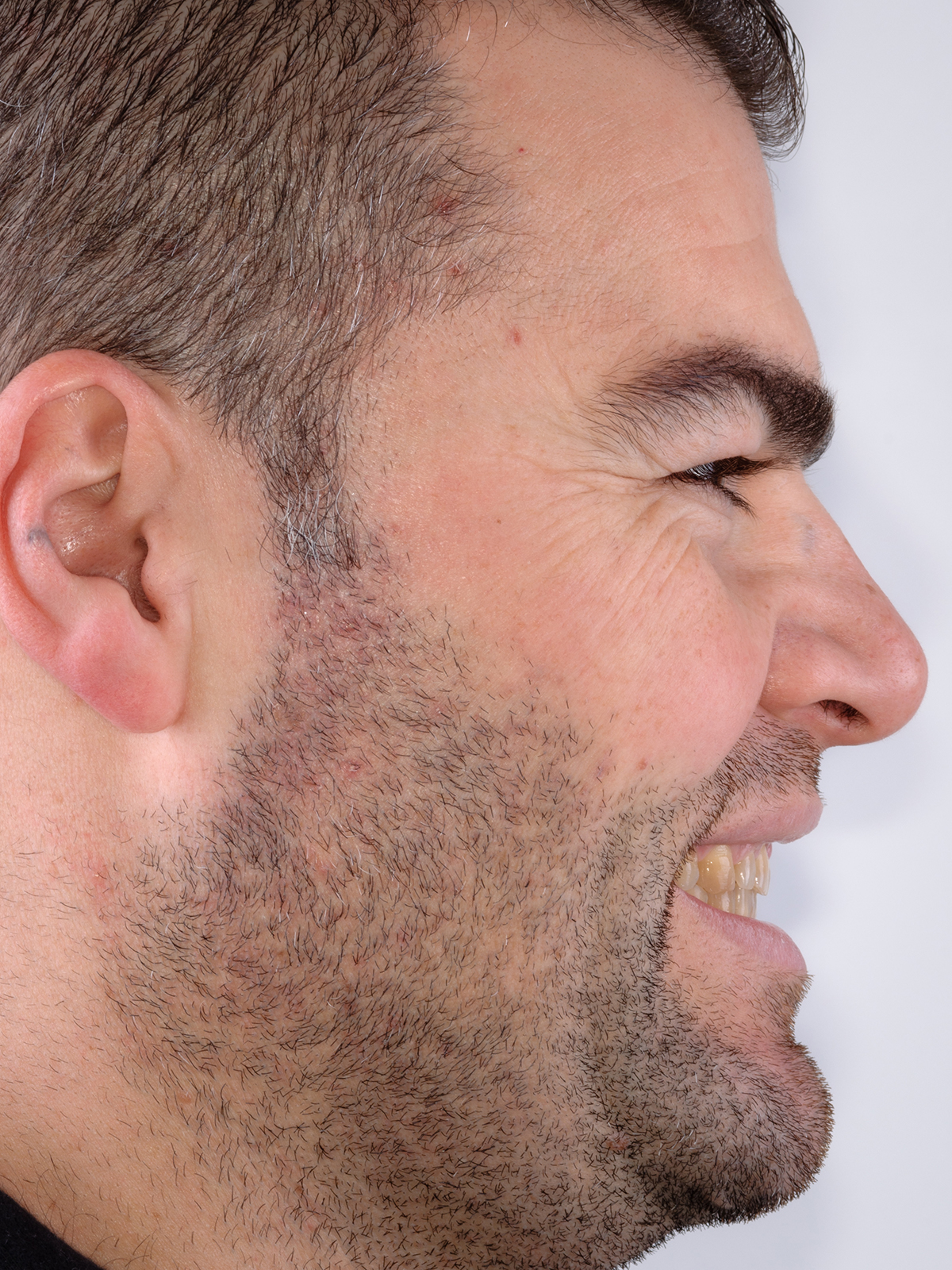
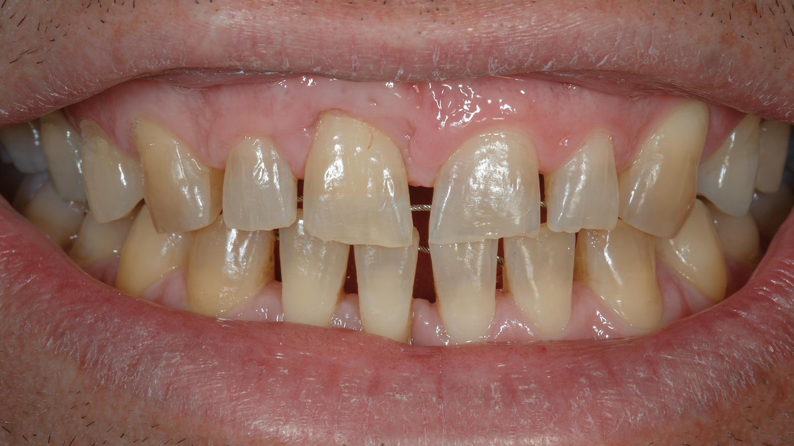
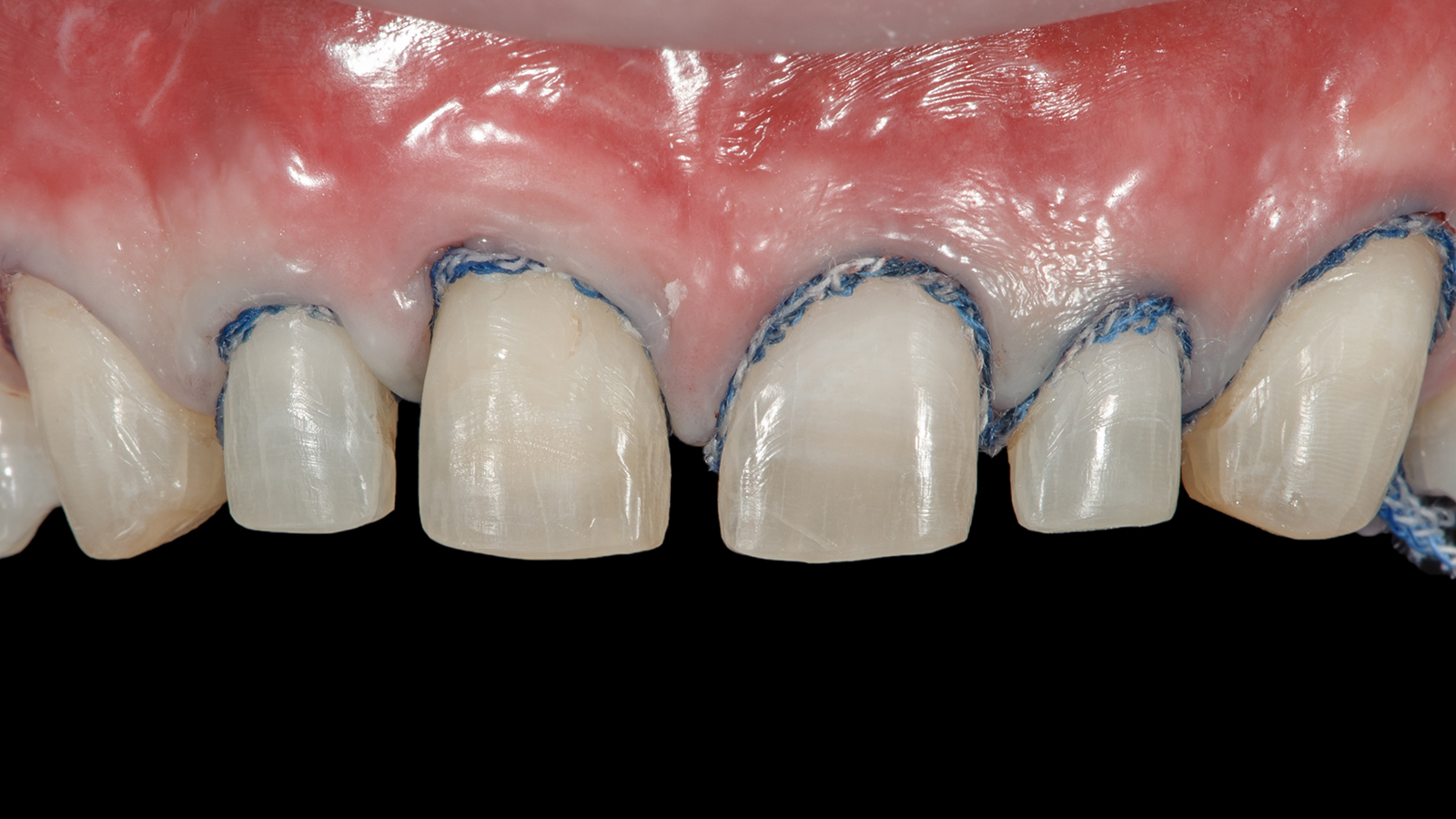
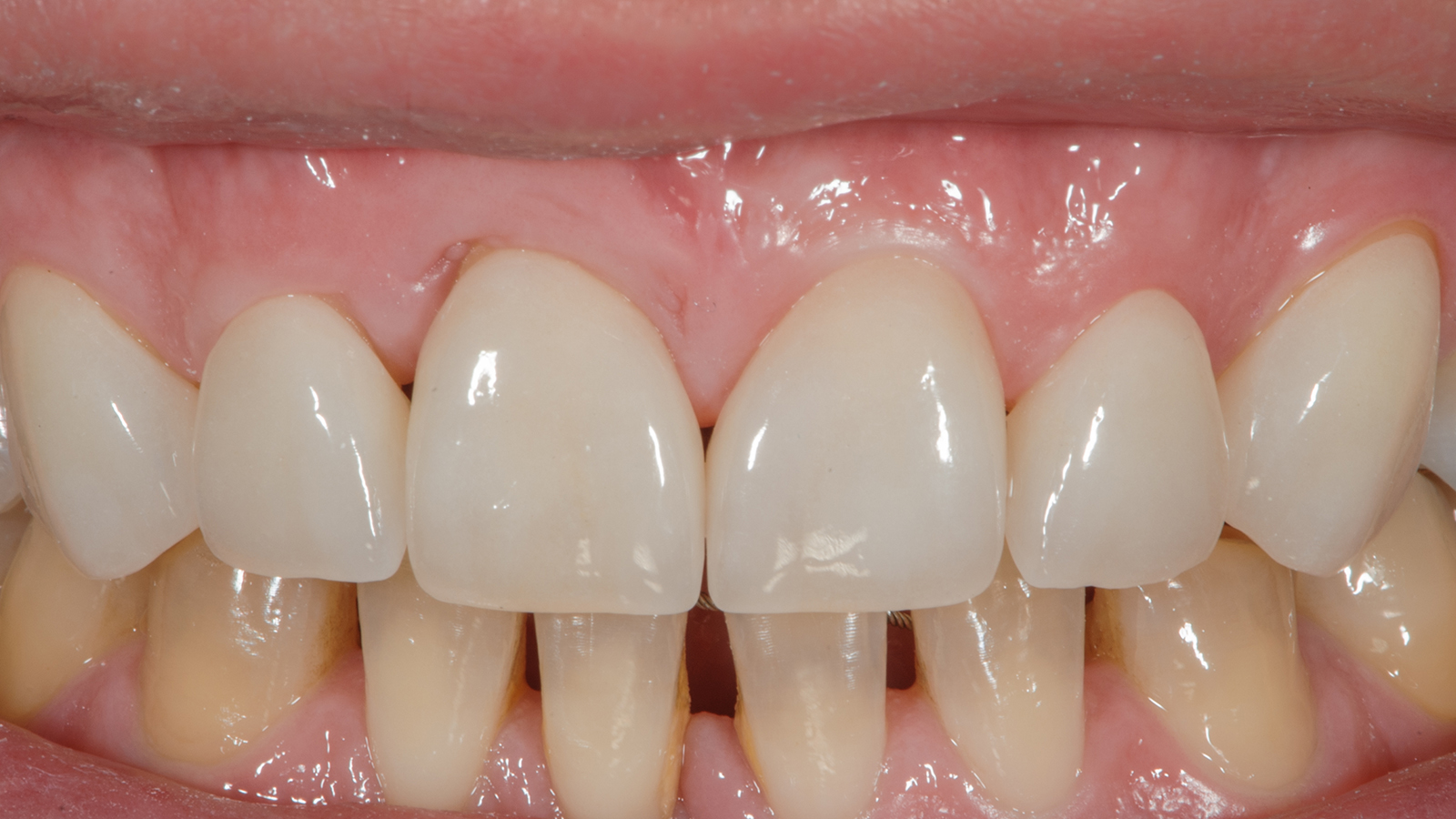
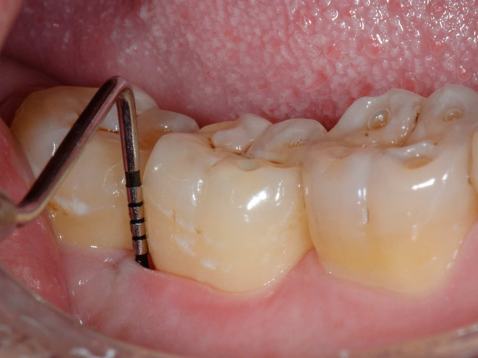
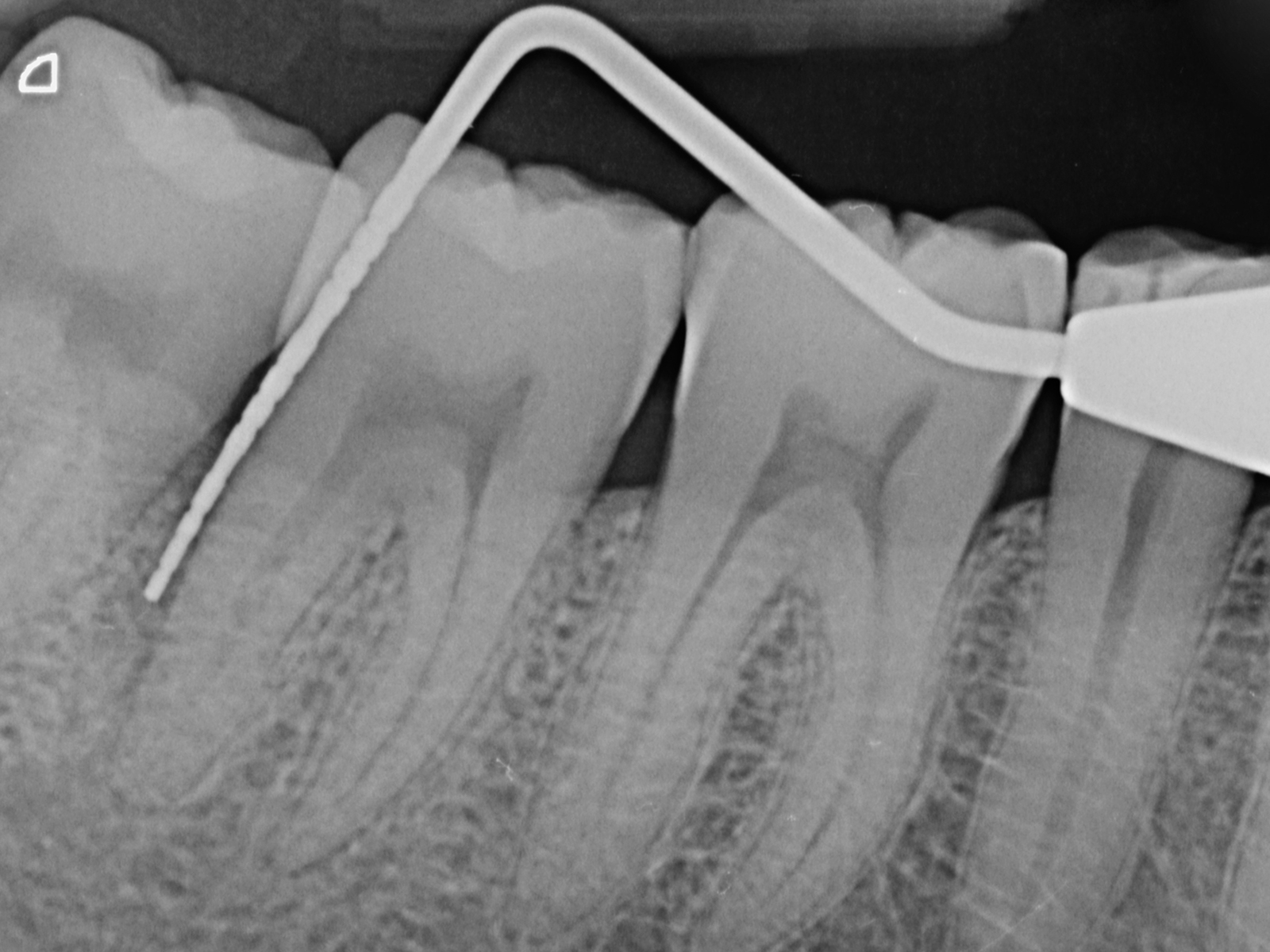
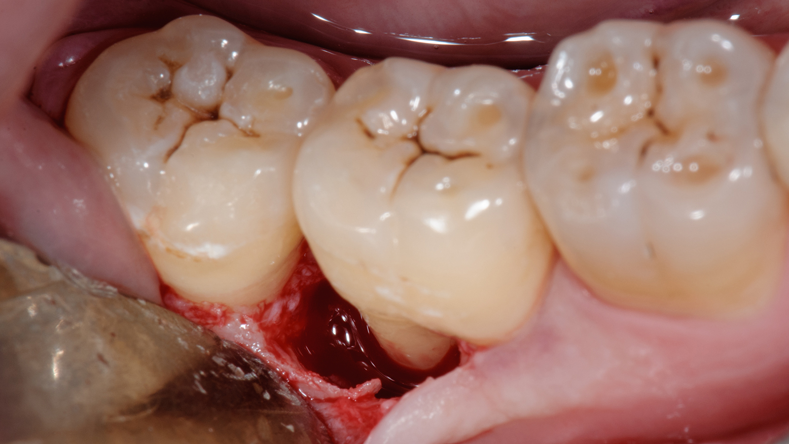
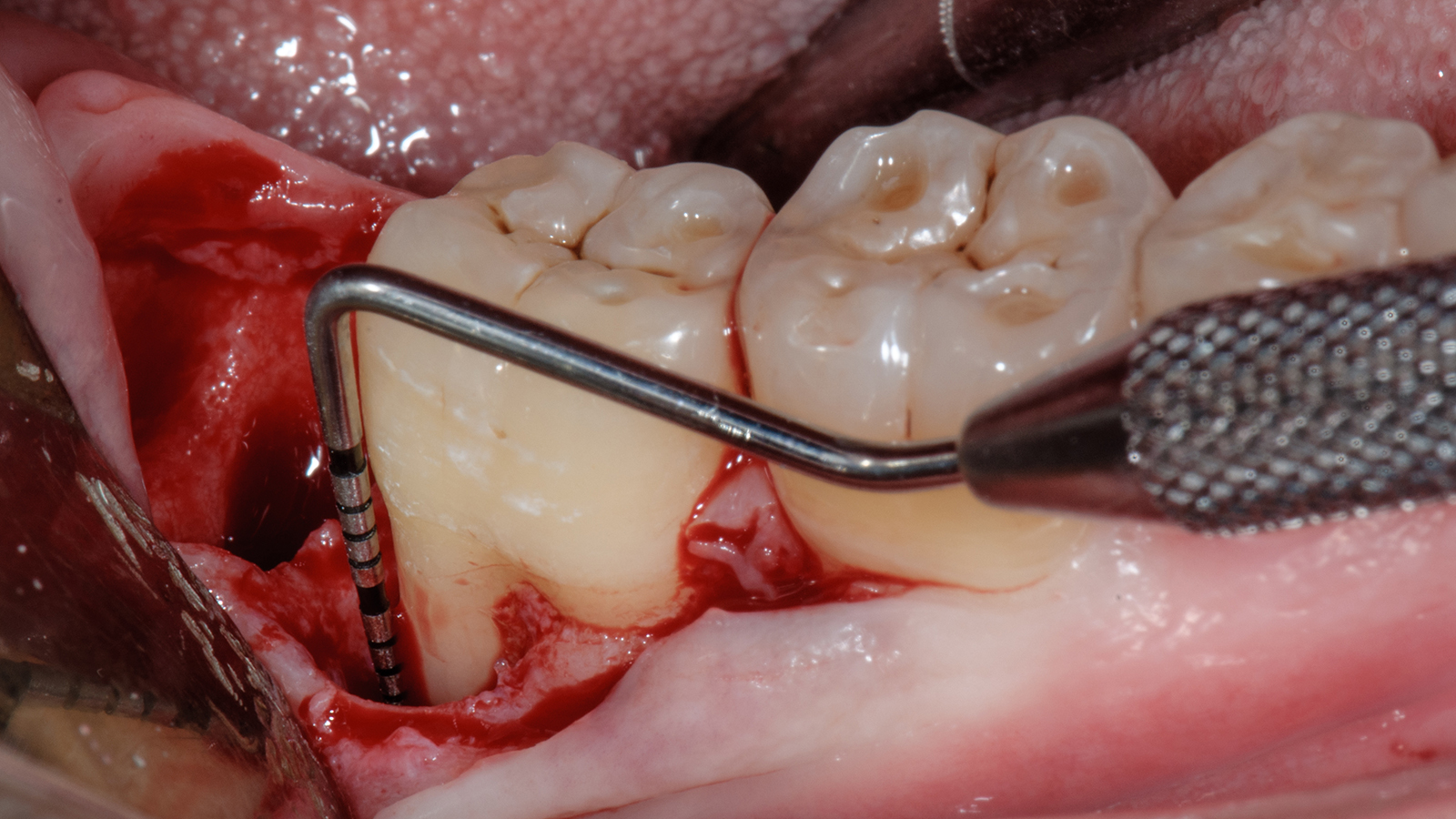
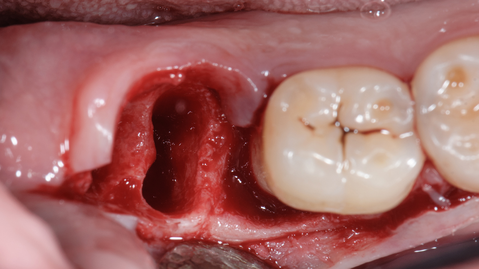
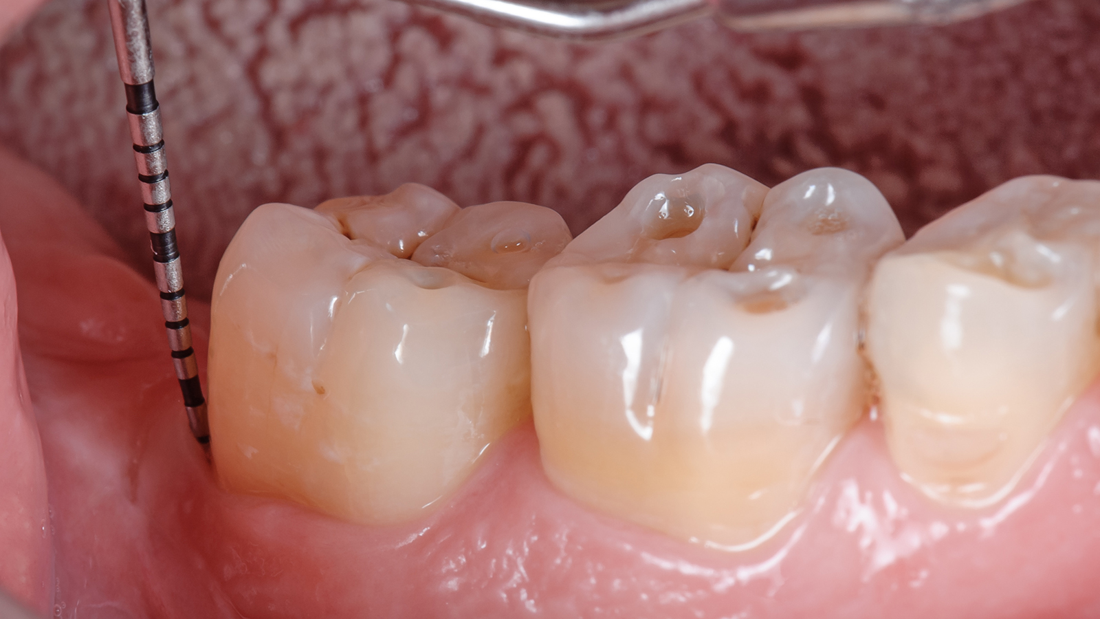
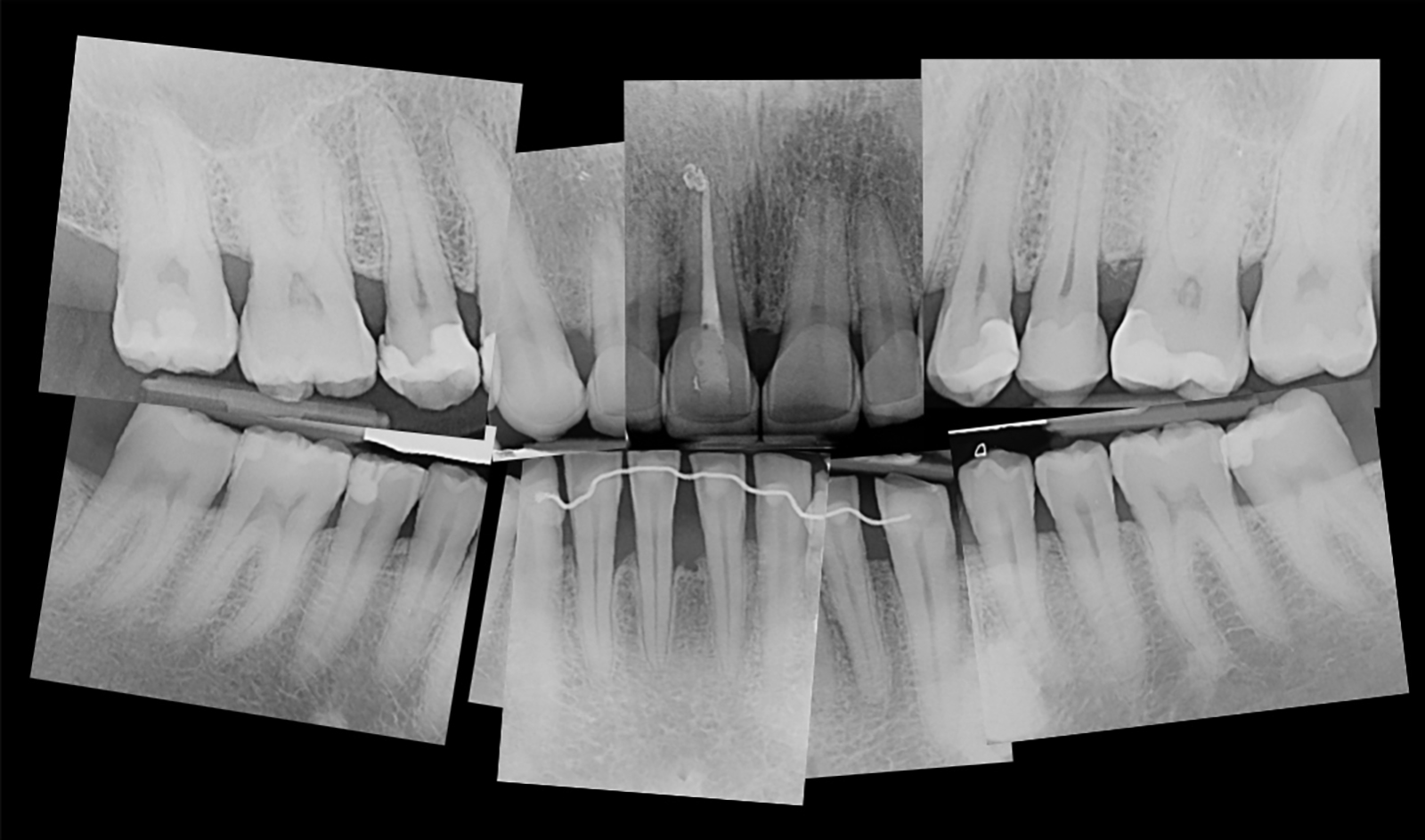
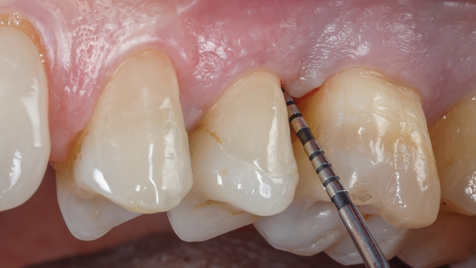
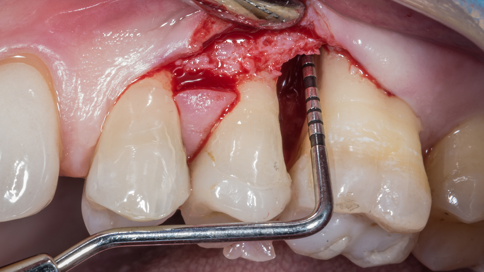
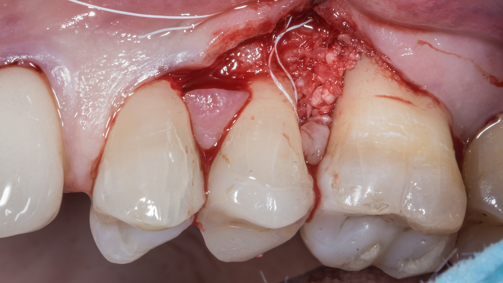
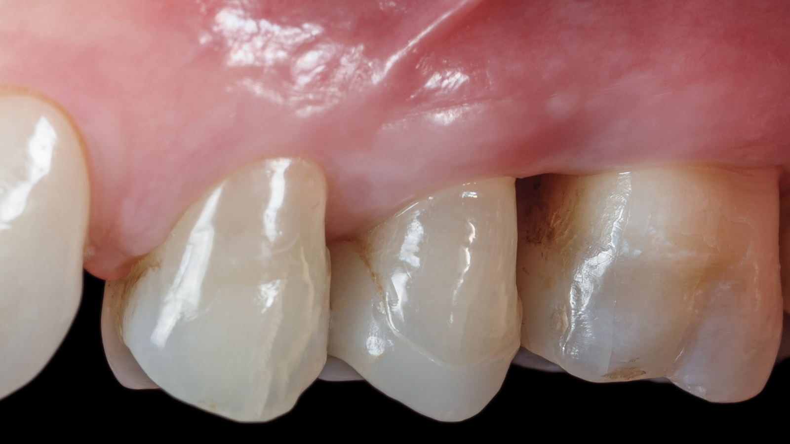
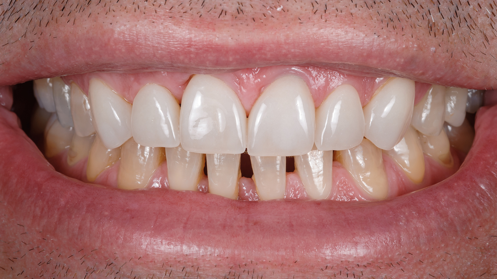
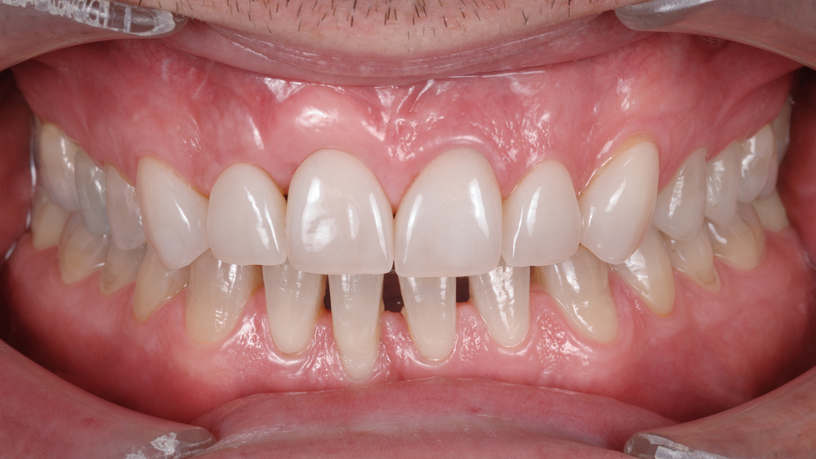
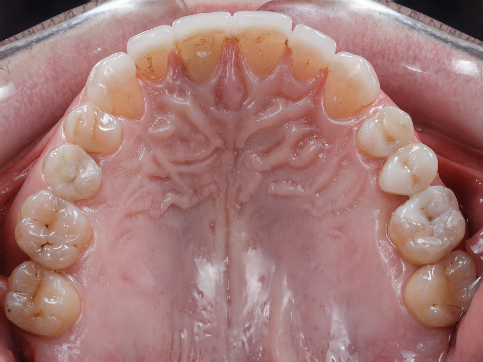
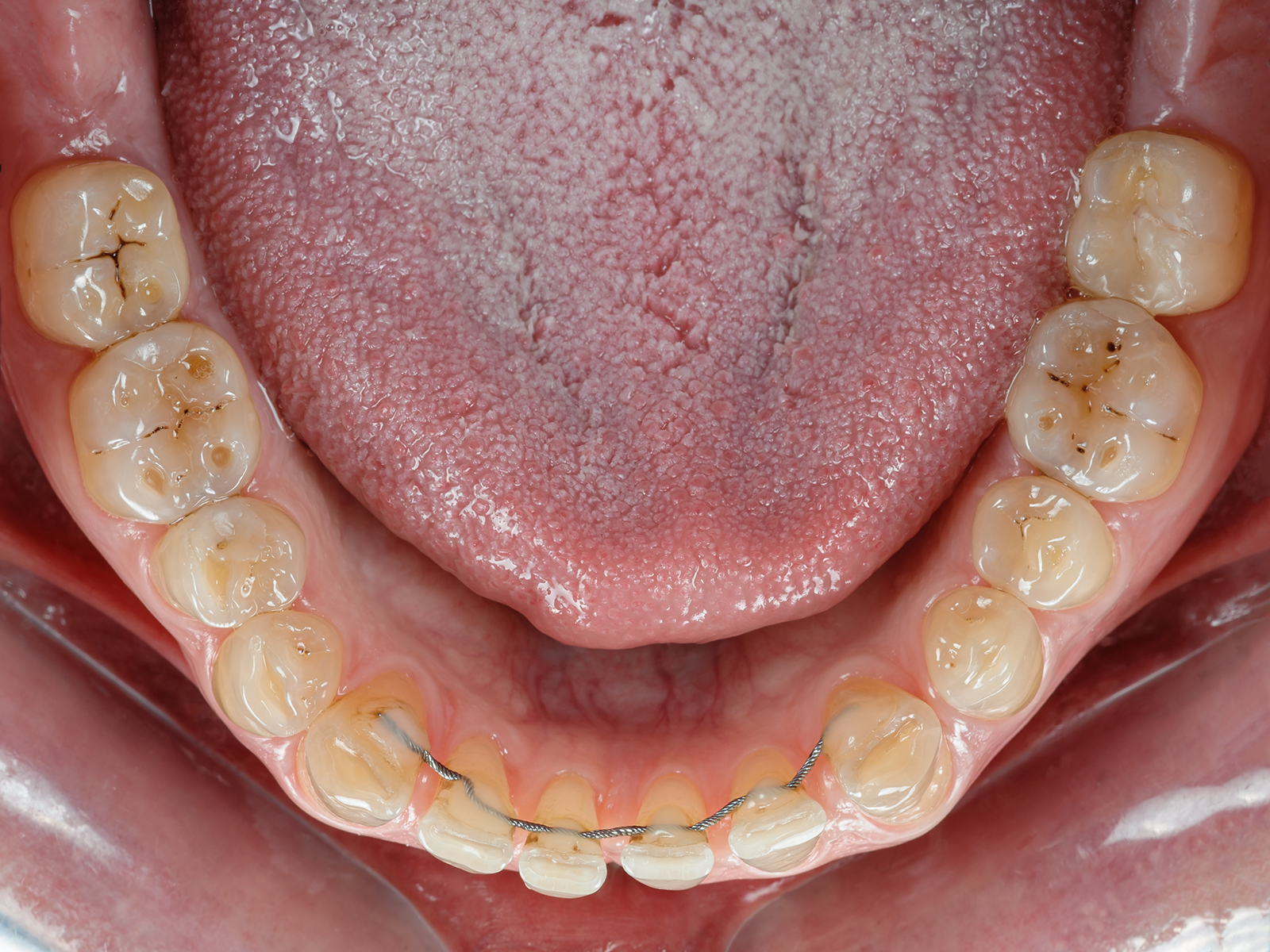
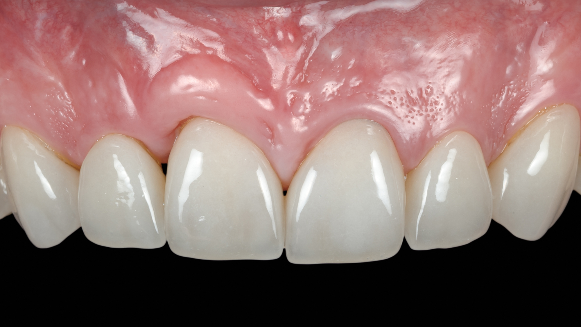
The patient presented with the chief complaint of pain and suppuration on tooth no. 8. He expressed that he had been previously treated for periodontal disease and had undergone bone regeneration surgery. He reported having regular brushing habits and consistent use of dental floss, interproximal brushes, and oral rinses.

Initial full face
Initial full face
Initial profile
Initial profile

Medical History

The patient does not smoke but drinks a glass of wine daily and takes Lansoprazol for heartburn and reflux. The patient reports no other medical issues.

Diagnostic Findings

Extraoral/Facial

  • Normal facial symmetry.
  • Centered midline.
  • Brachycephalic patient presenting a “gummy” smile showing more than 3 mm at maximum display.

TMJ/Mandibular Range of Motion/Muscles of Mastication and Facial Expression

  • Healthy TMJs.
  • Normal range of motion.
  • No history of clicking, pain or locking.
  • No muscle pain.

Intraoral Findings

Dental

  • Missing teeth nos. 1 & 16.
  • Teeth nos. 2, 4, 12 & 14 have amalgam restorations. Teeth nos. 5 & 13 have composite restorations.
  • Thick gingival phenotype.
  • Anterior teeth attrition.
  • Malposition with multiple diastemas in the anterior area.

Periodontal

  • All teeth except no. 8 have periodontal pocket depths between 3-5 mms.
  • Tooth no. 8 has distal pocket depth of 6 mms and buccal pocket depth 14 mms.
  • Tooth no. 7 has mesial pocket depth of 6 mms.
  • Suppuration around tooth no. 8.
Initial situation
Initial situation
Post-scaling probing

Occlusal Notes

  • Centric relation coincident with maximum intercuspation position.
  • Class I molar and canine relationships.
  • 2 mm overbite and 1 mm overjet.
  • Mild wear of anterior teeth secondary to protrusive bruxism movements.
  • No posterior excursive interferences.

Radiographic Analysis

Initial cephalometric radiograph
  • Mandibular third molars present.
  • Maxillary third molars missing.
  • Brachyfacial pattern.
  • Teeth with long roots and healthy periodontium.
Initial panoramic radiograph
Pretreatment FMX
Pretreatment FMX
Initial maximum intercuspal position
Initial maxillary occlusal view
Initial mandibular occlusal view

Diagnosis and Prognosis

  • Bruxism and mild attrition.
  • Stage II periodontal disease in most of the patient’s mouth and stage III in maxillary and mandibular posterior segments and teeth nos. 7 & 8.
  • Periodontal vertical defect on facial and distal of tooth no. 8 which has compromised the mesial of tooth no. 7.
  • Dental and skeletal class I.
  • Diastemas and mild malposition.
  • Good prognosis for all teeth except nos. 7 & 8.

Summary of Concerns

  • How do we treat the large vertical defect on the facial and distal of tooth no. 8 which affects the mesial bone of tooth no. 7?
  • Can and should tooth no. 8 be saved?
  • If dental implant replacement is considered, what would be the treatment sequencing?
  • If orthodontic treatment is considered, what would be the goals, objectives and sequencing?
  • How do we deal with the teeth malposition?

Proposed Treatment Plan

Options

  1. Extraction of tooth no. 8 and GBR (guided bone regeneration) plus dental implant placement. Followed by orthodontics and restorative treatment.
  2. GTR (guided tissue regeneration) and reevaluation prior to orthodontic and restorative treatment.

Phase I: Diagnostic Wax-up

  • For the initial case study, clinical and periodontal chartings, photos, full mouth series and panoramic radiographs will be obtained.

Phase II: Periodontal Therapy

  • Oral hygiene review with improvement in plaque control habits.
  • Monitor for gingival tissue improvement around tooth no. 8.

Phase III: Regenerative Surgery

  • GTR of the periodontal defect involving teeth nos. 7 and 8 utilizing a PTFE-e membrane.

Phase IV: Orthodontic Treatment

  • Reposition and realign teeth in both arches.

Phase V: Definitive Maxillary Restorative Treatment

  • Restore teeth nos. 6-11 with feldspathic porcelain veneers. The patient did not wish to restore the lower teeth.
  • Replace the worn amalgams with ceramic inlays and composites.

Phase VI: Maintenance

  • Oral hygiene maintenance.
  • Restorative and orthodontic maintenance.

Phase VII: Reevaluation

  • Reevaluation of the goals achieved and determine the need for any further treatment. Reevaluate the need for further periodontal treatment depending on the residual periodontal pocket depths.
V=veneer C/I=ceramic inlay © Seattle Study Club Journal

Active Clinical Treatment

Review of Treatment Goals

The patient’s primary objective was to keep tooth no. 8 and eliminate the pain and suppuration around it.

Phase I: Diagnostic Wax-up

Photographs, panoramic and full mouth intraoral radiographs, diagnostic casts mounted in CR (centric relation), and clinical and periodontal chartings were obtained.

Phase II: Periodontal Treatment

Overall periodontal health would be improved with root planing, scaling and oral hygiene review.

Phase III: Regenerative Surgery

To correct defects around teeth nos. 7 and 8, regenerative surgery was performed using a PTFE-e membrane (GORE-TEXÒ Regenerative Membrane Titanium Reinforced) fixed with two tacks and PTFE sutures. The biomaterial used consisted of autogenous bone harvested from the oblique line of the mandible. The periodontal defect was thoroughly cleaned, reaching almost the apex of tooth no. 8. A connective tissue graft was harvested from the palate and sutured on the buccal and proximal of tooth no. 8 to correct the gingival recession which appeared after the scaling. The PTFE-e membrane and the tacks were removed approximately one year later. During the second surgery, a fistula was detected on the buccal area of the regenerated bone. Tooth no. 8 was diagnosed as necrotic, and endodontic treatment was immediately performed. An additional connective tissue graft was harvested from the palate to compensate for the lack of volume due to buccal bone resorption caused by the fistula.

First Surgery

Second Surgery

Endodontic Treatment

Phase IV: Periodontal Control

While tooth no. 8 was being monitored for healing, several hygiene appointments were completed to maintain gingival health.

Tooth no. 8 follow-up

Phase V: Orthodontic Treatment

The orthodontic treatment started one year later to correct the malposition in the anterior segment. This treatment lasted one year.  During this time composite restorations of teeth nos. 21 & 28 were completed and tooth no. 17 was extracted to help maintain the periodontal health of tooth no. 18.

Orthodontic treatment
Post-orthodontic treatment full face
Post-orthodontic treatment profile
Post-orthodontic smile

Phase VI: Bleaching & Definitive Maxillary Restorative Treatment

Before restoring the anterior segment, intraoral bleaching was done. After the wax-up and mock-up was performed to assess esthetics and function, teeth nos. 6-11 were restored with feldspathic veneers.

The treatment was finalized with a night guard to protect the restorations.

Mock-up
Mock-up
Minimally invasive preparations
Teeth nos. 6-11 veneers

Phase VII: Maintenance

Periodontal maintenance was performed periodically, however several maintenance appointments were missed by the patient. In 2015, a periodontal defect was detected in the distal of tooth no. 31. A GTR procedure was performed and tooth no. 32 was extracted to avoid food impaction and to improve the long-term prognosis of tooth no. 31.

Tooth no. 31 probing
Tooth no. 31 radiograph
Tooth no. 31 GTR
Tooth no. 31 GTR
Tooth no. 31 GTR
Tooth no. 31 GTR
Tooth no. 31 GTR
Tooth no. 31 GTR
Tooth no. 31 GTR follow-up

In 2016, resective periodontal surgeries on teeth nos. 2, 3, 14 & 15 were completed to reduce the depth of periodontal pockets, facilitating hygiene.

Resective periodontal surgery
Resective periodontal surgery follow-up

In 2021, a regenerative periodontal surgery was completed with Endogain and MinerOss 0,5 for teeth nos. 13 & 14.

Regenerative periodontal surgery
Regenerative periodontal surgery follow-up

Regular oral hygiene, restorative and orthodontic maintenance recall appointments continue.

Maintenance smile
Maintenance smile
Maintenance maximum intercuspal position
Maintenance full face
Maintenance profile
Maintenance profile
Final maxillary occlusal view
Final mandibular occlusal view
Final veneers

Commentary

Treating this patient early in my career proved to be lengthy and quite challenging. Even with more years of experience under my belt, I believe that this would still be difficult and perplexing for most clinicians.

In retrospect, I would probably not change my approach to this case. I spoke to the patient honestly and explained the anatomy of the bone defect affecting both teeth nos. 7 and 8. I explained that the most straightforward option would be to extract tooth no. 8 but that vertical bone regeneration would be almost impossible due to the lack of a bone peak on the mesial of tooth no. 7. Orthodontic extrusion would be then needed to reposition the bone peak coronally, but the 6 mm periodontal bone defect would force us to extrude tooth no. 7 too much and he would end up requiring endodontic treatment and crown lengthening on at least the distal of the tooth. All of that would compromise the crown-to-root ratio of tooth no. 7 and as a result, its long-term predictability. So, we discarded that option.

Extracting both teeth nos. 7 and 8 (observing the coronal peaks on both sides of the defect) would have been a very aggressive approach for this young patient, involving multiple surgeries and a high “esthetic” risk. Uncertain papilla levels and volume and the patient’s “gummy” smile would cloud the predictability of the outcome.

It was decided to proceed with GTR of the 0-wall periodontal defect even knowing the high risks of a possible membrane exposure. The main reason to start with this was that we always had the other two options in case of failure. I used a CTG to allow for better membrane coverage and to correct the mild gingival recession present after the initial scaling. This was successful, but after removing the PTF-e membrane during the second stage surgery, it was evident that the large periodontal defect had compromised the pulp’s vitality. We completed the endodontic treatment during the same appointment and harvested another CTG to compensate for the lack of buccal bone volume.

Additional periodontal defects were treated over the subsequent years in part because the patient was less than compliant with recall appointments.

Even though there were some setbacks during treatment, the patient was happy with the final result. After 15 years, the treatment continues, the patient’s intraoral condition has improved, and the long-term prognosis of the teeth has changed for the better.