Case 13: Special Report 13 — Orthodontic Extrusion Prior to the Placement of Adjacent Implants for a More Esthetically Pleasing Smile

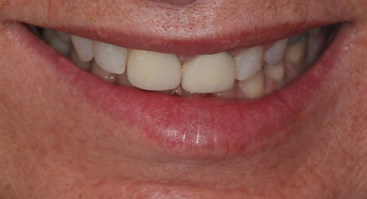
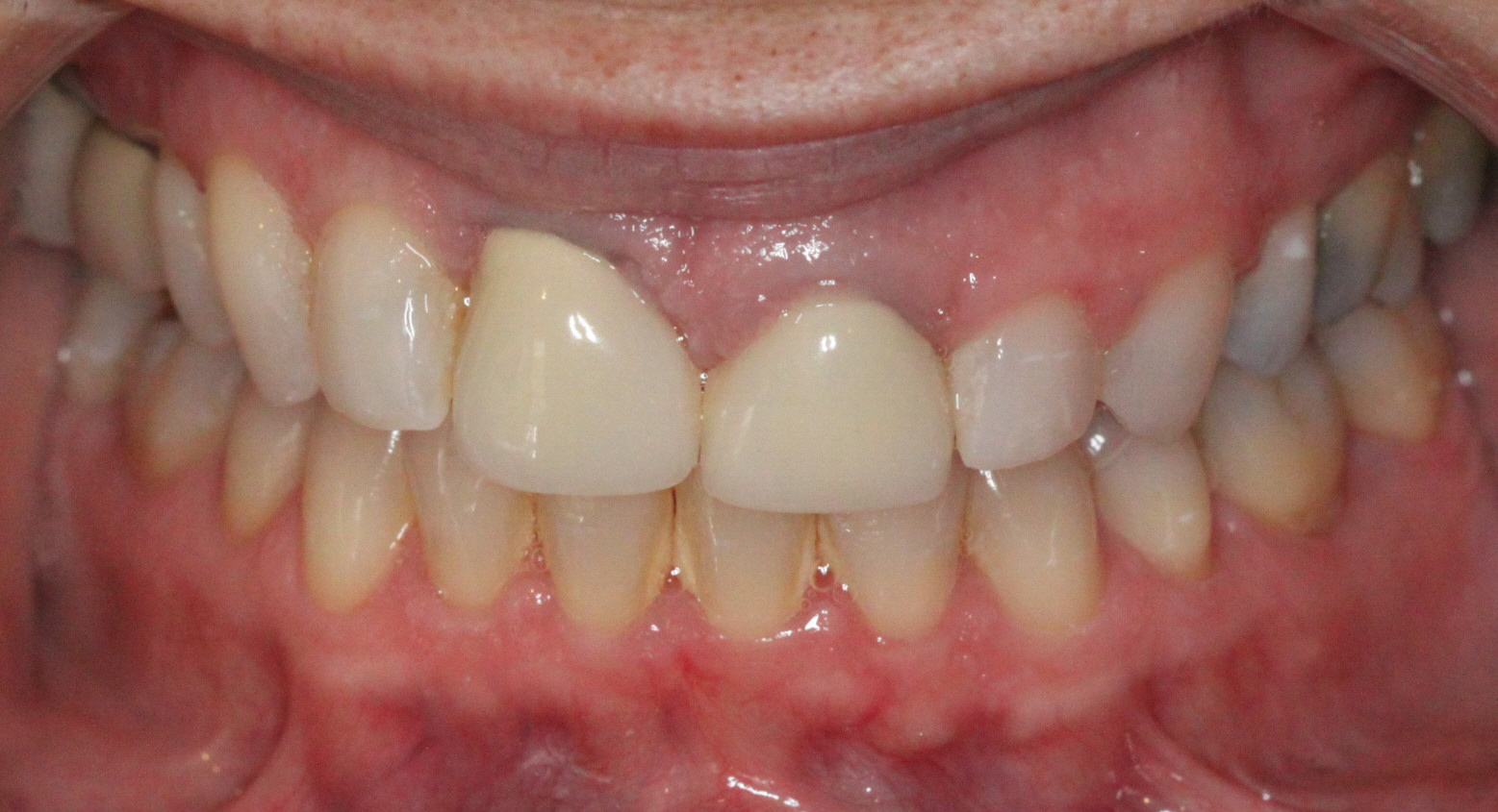
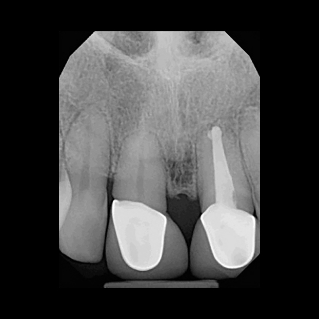
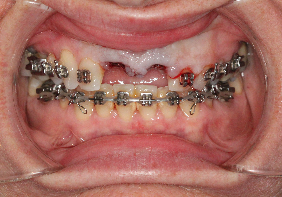
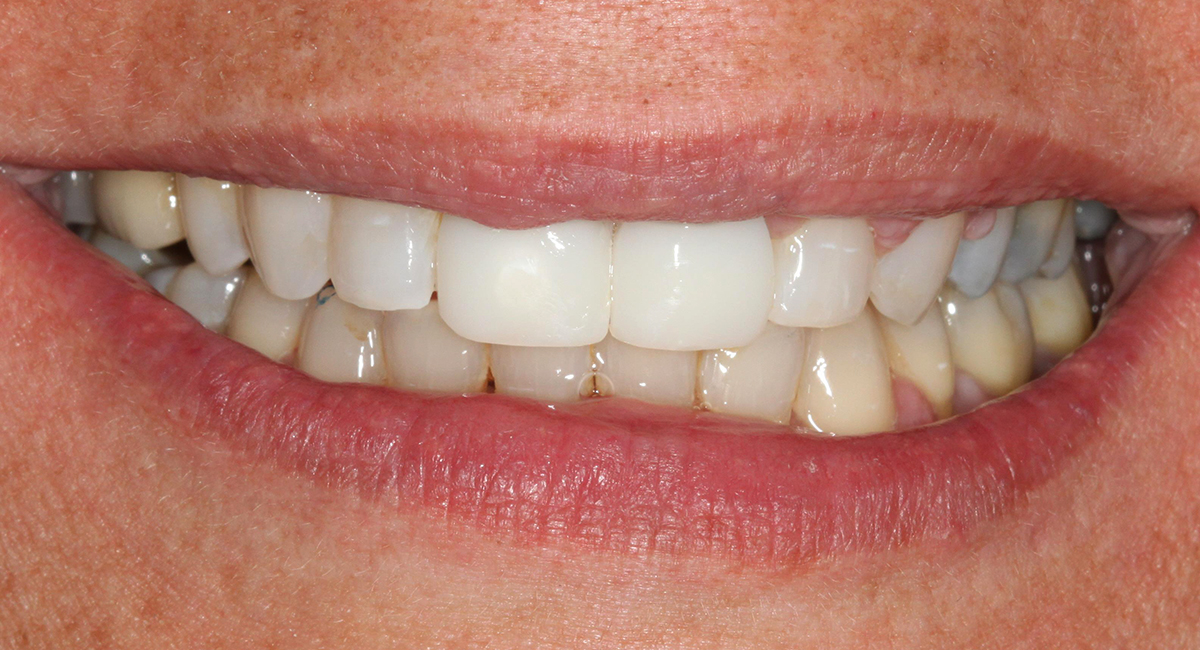
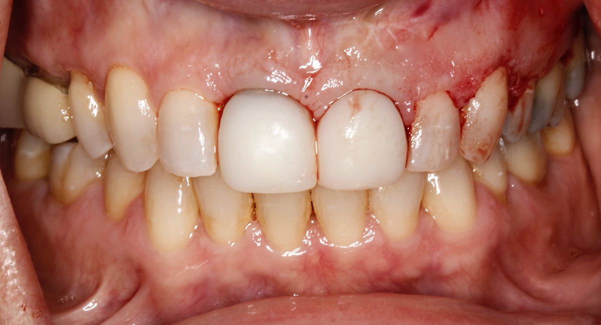
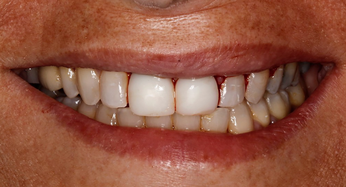
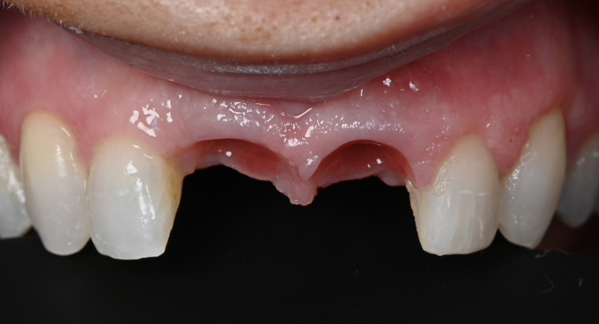
Implant restoration in the esthetic zone often requires well-coordinated interdisciplinary care in order to achieve ideal treatment results. This is especially the case when implants are planned to replace periodontally hopeless teeth. When teeth are extracted, we can typically anticipate 2mm of facial recession. If implants are to be placed following the extraction of hopeless teeth with an already existing gingival height discrepancy, the discrepancy will likely be exacerbated.

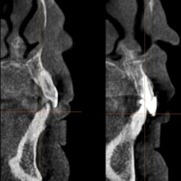
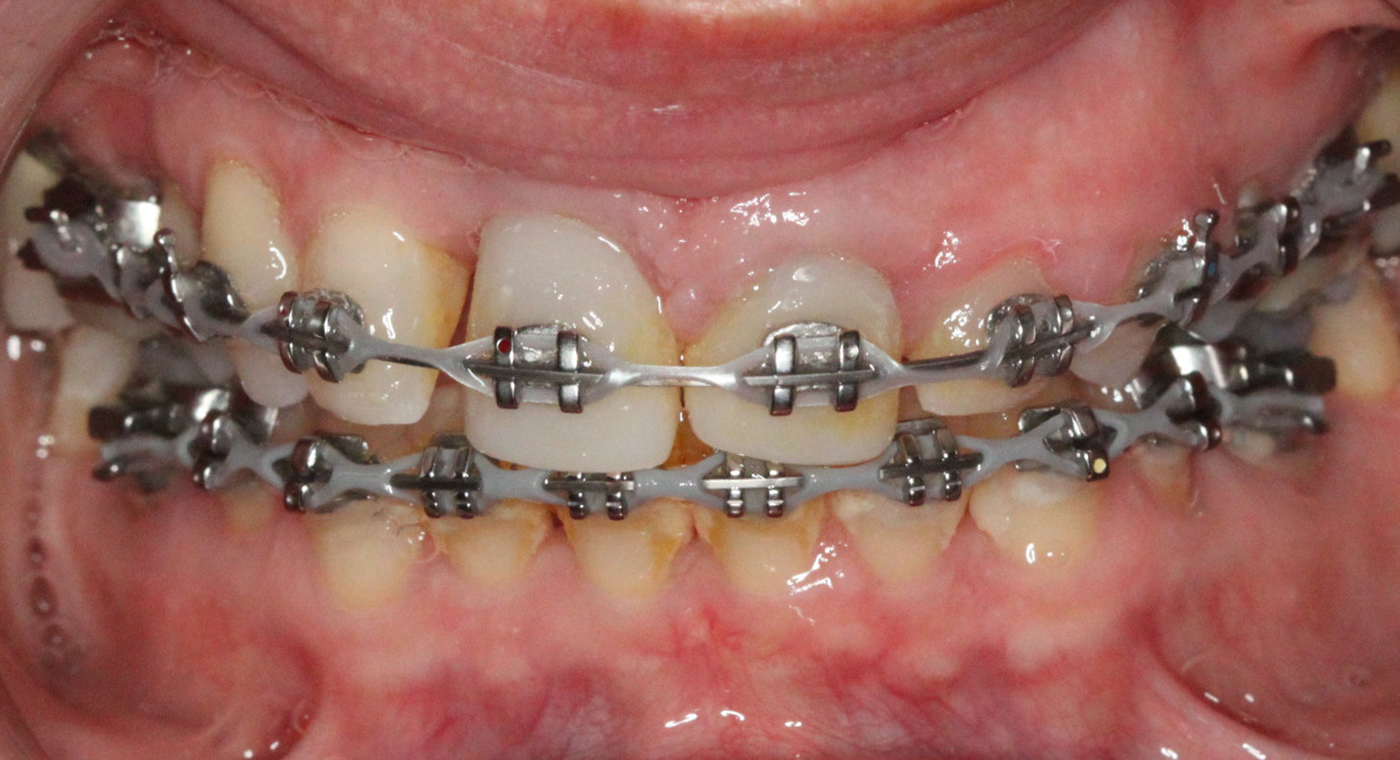
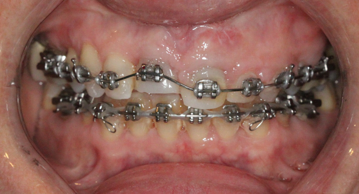
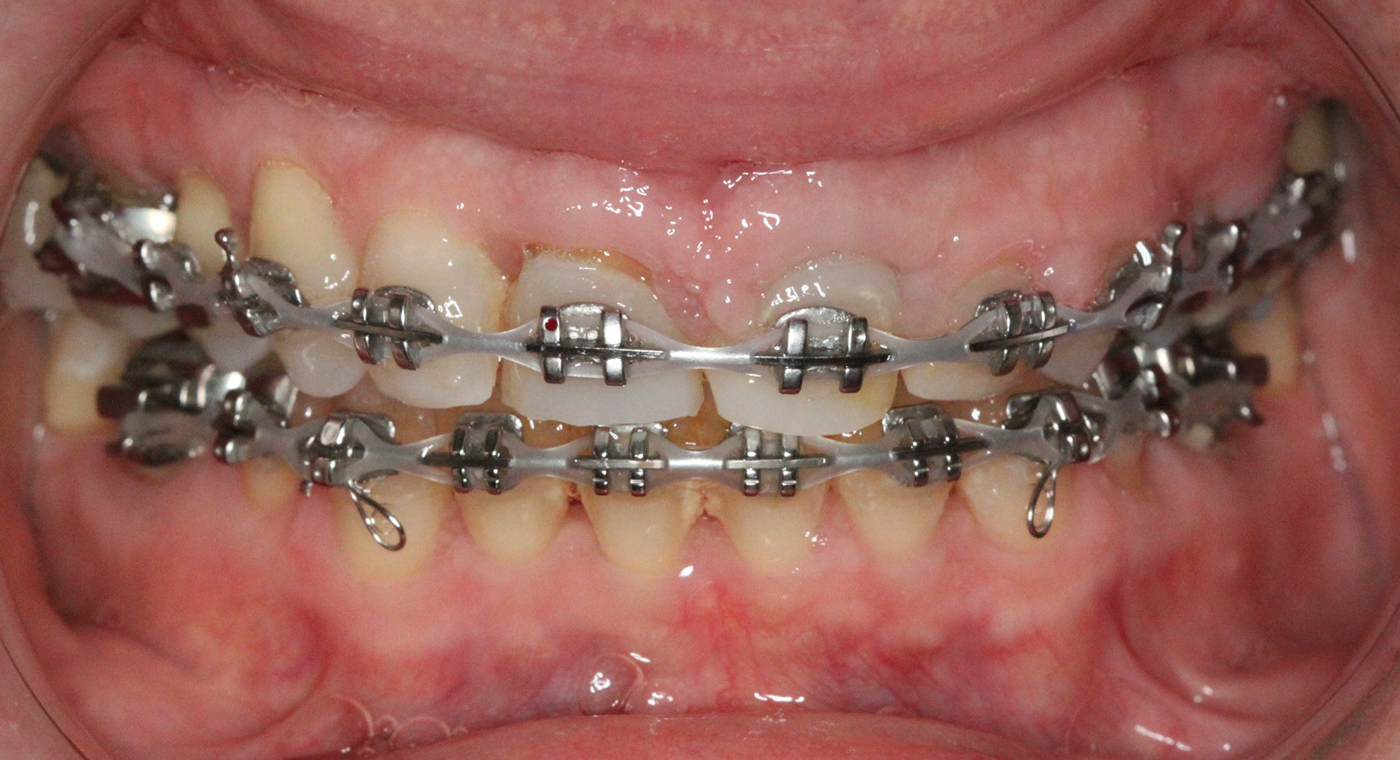
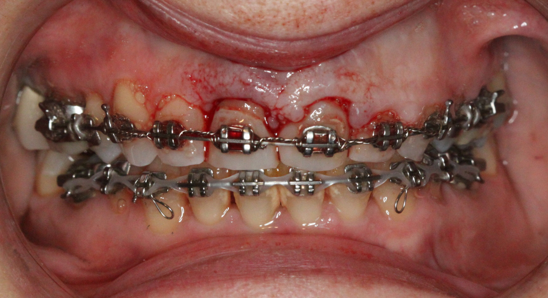
Extraction after forced eruption can allow for a much more favorable implant site compared with extraction alone. This is possible because the tension applied to the periodontal ligament during orthodontic tooth movement stimulates osteoblastic activity to induce new bone formation. As the tooth moves coronally during extrusion, soft tissue and bone attached to the periodontal fibers migrate in the same direction. As a result, forced eruption can be used to enhance the quality and quantity of both hard and soft tissue of future implant sites.

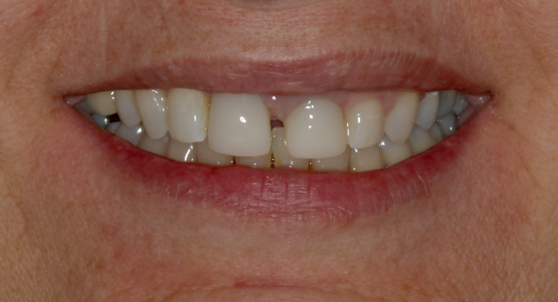
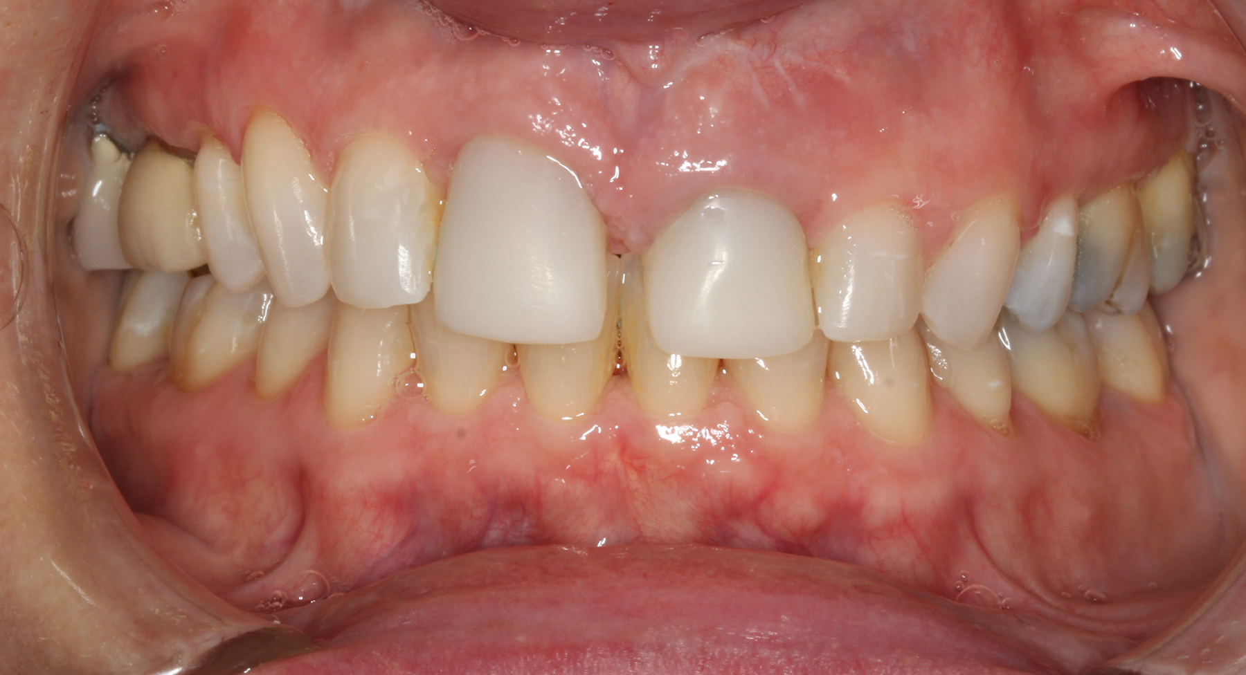
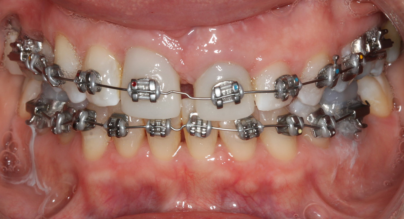
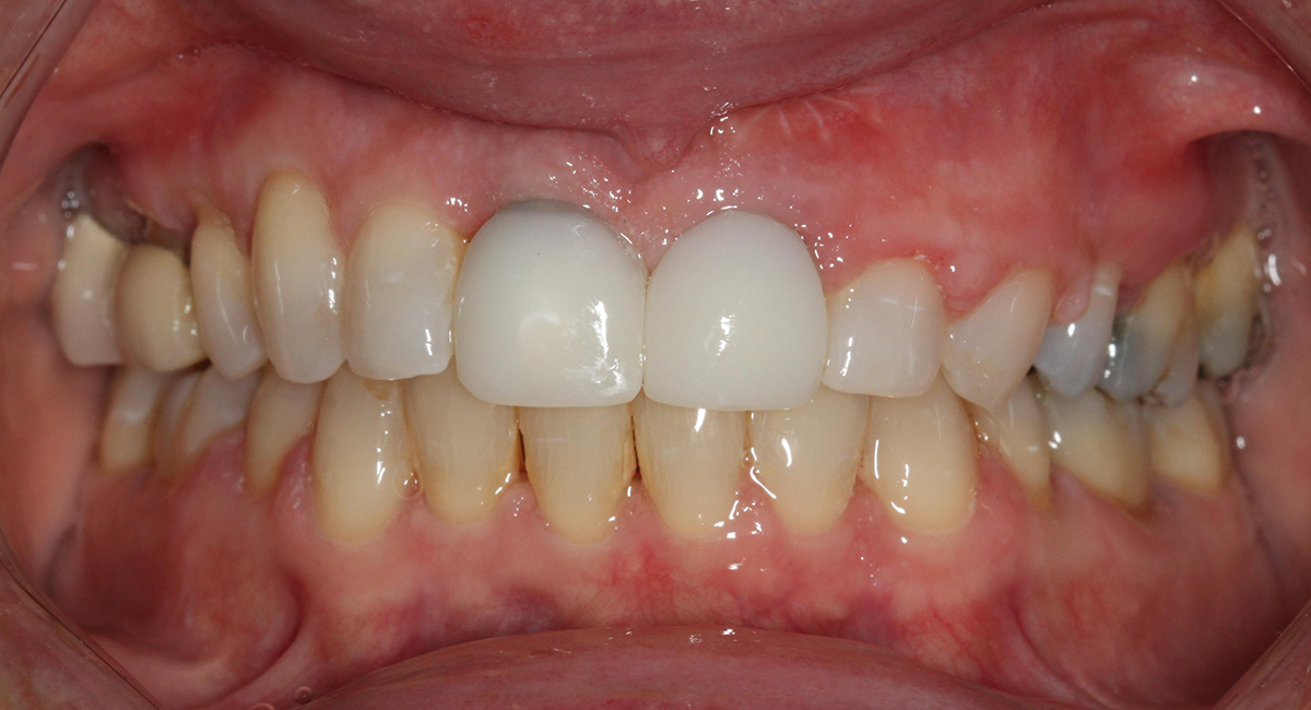
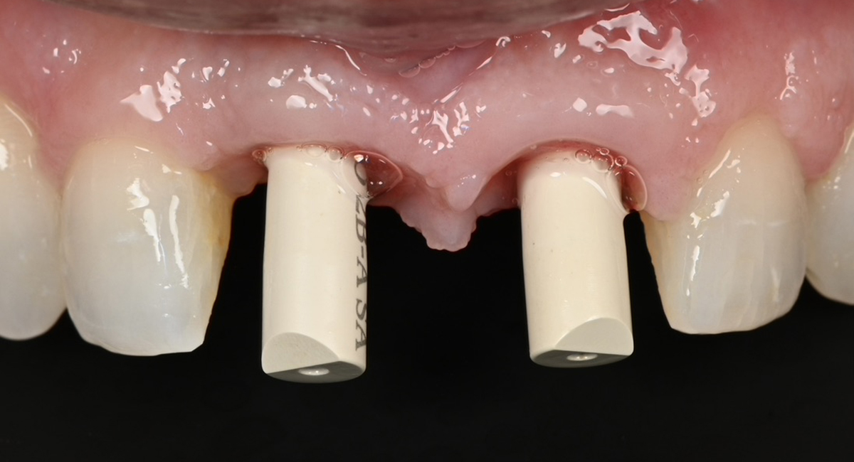
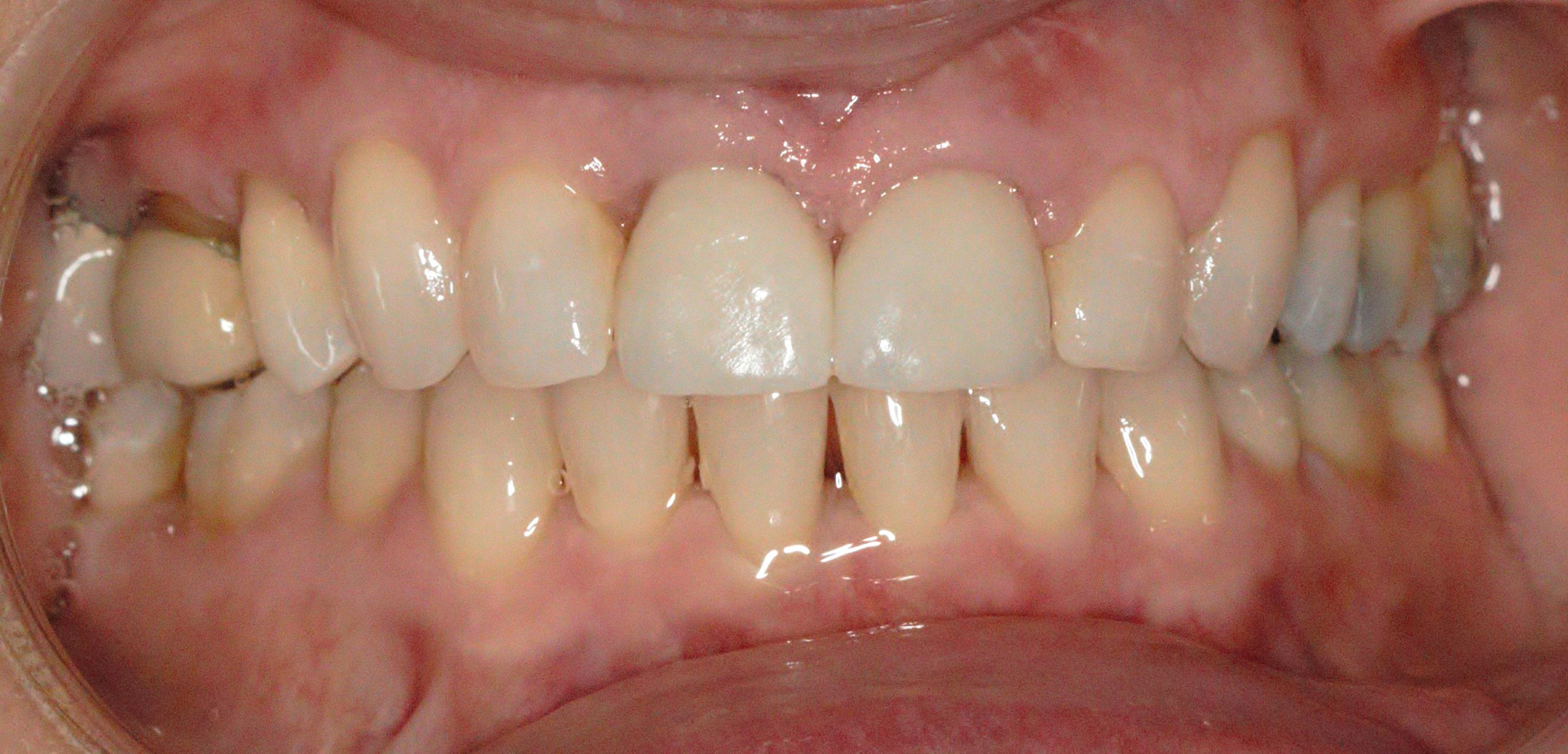
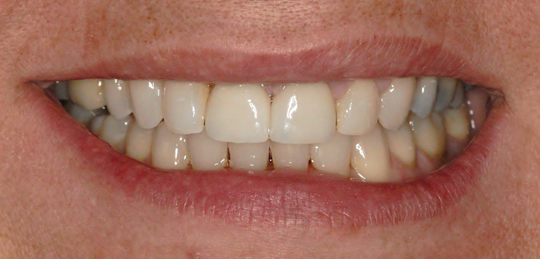
The special report presented below illustrates how orthodontic extrusion was successfully used to improve the periodontal architecture prior to the placement of two adjacent implants in the esthetic zone and thus allowed for a more ideal restorative result.

Dental History

A 58-year-old patient presented to us after completion of clear aligner treatment with her general dentist. Her medical history included a heart murmur, mitral valve prolapse, hemophilia, high blood pressure, kidney problems, and anxiety. She was taking Bystolic, Norvasc, Lexapro, and premedicating with Amoxicillin.

The patient described a history of trauma to her anterior teeth as a child and had recently fractured her maxillary left central incisor at the gingival margin. While content with her alignment and occlusion, both patient and dentist desired closure of space between her maxillary central incisors, even gingival margins, and a more esthetically pleasing smile.  Achievement of these goals required a true interdisciplinary approach.

Data Acquisition

  • Medical and dental history
  • Comprehensive clinical (extra-oral, intraoral, dental, periodontal, functional, esthetic) and radiological exams
  • Intraoral scans of both arches (iTero, Align Technology, Arizona, United States)
Initial full face, smiling
Initial full face in repose
Initial smile
Initial smile
Initial maximum intercuspation
Initial maxillary occlusal view
Initial mandibular occlusal view
Initial sagittal CBCT images nos. 8 and 9
Initial periapical image of nos. 8 and 9

Diagnosis

Based on the clinical and radiological findings, the following diagnoses were determined:

  • Extraoral: Straight profile, short lower face height, nose slightly to the right, lower lip asymmetrical, chin button off to the right
  • Intraoral: Excessive buccal corridors right and left, occlusal cant upwards to the right
  • Dental: Porcelain-fused-to-metal crowns nos. 2, 3, 8, and 9, Angle Class I canines and molars, lower midline 1mm to the right of upper midline, uneven tooth length of all maxillary anteriors: 6mm (no. 5); 10mm (no. 6); 9mm (no. 7); 11mm (no. 8); 9mm (no. 9); 7mm (no. 10); 8mm (no. 11); 6mm (no. 12)
  • Periodontal: Uneven gingival margins on the upper, altered passive eruption of nos. 10 and 11, even gingival margins on the lower, gingival health on a reduced periodontium
  • Radiographic: thin facial bone nos. 8 and 9, poor crown-to-root ratio no. 8, apical root resorption no. 8, previous endodontic treatment and apicoectomy of no. 9, no evidence of decay
  • Prosthetic: Crowns nos. 8 and 9 wider than ideal
  • Esthetic: Compromised esthetics

Based on the diagnosis, the prognosis of nos. 8 and 9 was determined to be poor and these teeth were treatment planned for extraction and replacement with adjacent implants. Prior to implant placement, we decided to use orthodontic extrusion to create even gingival margins for the central incisors and allow for implant site development.

Due to the apical level of the interproximal bone between nos. 7 and 8, the papilla between these teeth was more apically positioned than ideal. We opted against extruding no. 7 to correct the papilla as doing so would have placed no. 7 in a non-ideal position.

Active Clinical Treatment

Phase I: Pre-Orthodontic Restorative Treatment

Existing crowns on nos. 8 and 9 were removed and replaced with provisional crowns of ideal width.

Restoration of maxillary centrals with provisional crowns of ideal width

Phase II: Comprehensive Orthodontic Treatment

Fixed orthodontic appliances were then placed on both upper and lower arches. After completion of leveling and aligning, all spaces were closed.

Placement of fixed orthodontic appliances
Orthodontic space closure

Prior to the extrusion of no. 8, the incisal edge of this tooth was trimmed. The bracket on this tooth was then apically repositioned to facilitate its eruption. The lingual surfaces of nos. 8 and 9 were reduced to allow for retraction of these teeth, to continue space closure, and to correct their inclination. Class III elastics were also used to achieve these goals.

Reposition of bracket on no. 8 to allow for orthodontic extrusion
Orthodontic extrusion completed

Phase III: Digital Implant Planning, Extraction and Implant Placement

Once the patient’s occlusion was stable and we had achieved even gingival margins of nos. 8 and 9, these teeth were extracted. Two implants (Nobel Biocare AG, Gothenberg, Sweden, active narrow platform, 3.5mm width, 13mm length) were placed on the same day using a digitally fabricated stent. Gingival hyperplasia noted interproximally between the implants can be attributed to the patient’s use of Norvasc, a calcium channel blocker for hypertension.

Digital treatment planning of implant placement nos. 8 and 9 – sagittal view
Digital treatment planning of implant placement nos. 8 and 9 – occlusal view
Digital treatment planning of implant stent – sagittal view
Digital treatment planning of implant stent – occlusal view
Surgical implant guide in place
Periapical radiograph after implant placement

Implants were placed subcrestally and 3mm tall healing caps were selected to allow for both access and for the extracted teeth to be altered and to be used as pontics.

Same day pontic placement was then completed using the crowns of the extracted teeth. These teeth were trimmed accordingly and then ligated to the patient’s upper archwire. Placement of ovate pontics in the extraction sockets allowed for the maintenance of the gingival architecture during implant integration. Teeth nos. 6-11 were splinted together for stability.

Periapical radiograph illustrating implants, healing caps, and pontics made with extracted teeth nos. 8 and 9
Implant placement – maxillary occlusal view
Implant placement – intraoral maximum intercuspation view
Placement of pontics using extracted teeth nos. 8 and 9 — maxillary occlusal view
Placement of pontics using extracted teeth nos. 8 and 9 — maximum intercuspal position

Phase IV: Implant Temporization and Esthetic Crown Lengthening

After allowing for an osteointegration period of four months, orthodontic appliances were removed and provisional implant crowns were placed on nos. 8 and 9 on the same day.

Three weeks later, esthetic crown lengthening was then performed on nos. 10 and 11 to correct the altered passive eruption of these teeth.

Six weeks after placement of the provisional implant crowns, an intraoral scan was taken in order to fabricate final implant crowns.

Provisional implant crowns nos. 8 and 9 – maximum intercuspal position
Provisional implant crowns nos. 8 and 9 – smile
Esthetic crown lengthening nos. 10 and 11 – maximum intercuspal position
Esthetic crown lengthening nos. 10 and 11 – smile
Gingival margins at intraoral scan for final implant crowns
Implant scan bodies in place

Phase IV: Definitive Restorative Treatment

Final implant crowns were then delivered for nos. 8 and 9. Due to the improved appearance of nos. 7 and 10 following orthodontic treatment and after esthetic crown lengthening of nos. 10 and 11, we decided against the need for bonding or veneers for these teeth. Additionally, although the height of the papilla in between nos. 7 and 8 decreased slightly following extraction, we expect it to return to its original height.

Definitive full face, smiling
Definitive full face in repose
Definitive maximum intercuspation
Definitive smile
Definitive smile

Commentary

This special report illustrates the successful transformation of a patient’s smile with minimally invasive dentistry. This was possible through the use of orthodontic treatment to improve the foundation on which two adjacent implants were placed, the use of esthetic crown lengthening to create a symmetrical smile, and restoration of the implants with crowns of ideal proportion.