Helen Roberts
Scientific Manager, 3Shape
This article is an excerpt from the ebook Because quality treatment matters to me —
What research and practitioners tell us about intraoral scanners and treatment quality.
Since their initial introduction to the market, clinicians have sought to verify intraoral scanning accuracy. Unfortunately, conducting a study with a tool (IO scanner) operated by a human hand can inherently present challenges. Besides possible human error when scanning, scan strategy can impact accuracy results significantly. Moreover, the patient will always be a variable and a reason most studies are conducted in-vitro.
The difference between a conventional impression and digital impression is that analog impression material records a space by using physical pressure. A digital impression documents anatomy by imaging the margin shape with an optical system.
Because of this, depending on the intraoral scanner, it may be difficult to scan challenging subgingival conditions. As a result, scan and preparation strategies need to be adjusted to meet these challenges.
The Dr Watt Method
An example of adapting to the technology is given by UK practitioner Dr Douglas Watt in a case study he presented.
Dr Watt found the biggest challenge during a clinical case “was scanning adjacent subgingival margins and getting a clear marginal definition all the way round the preparation.”
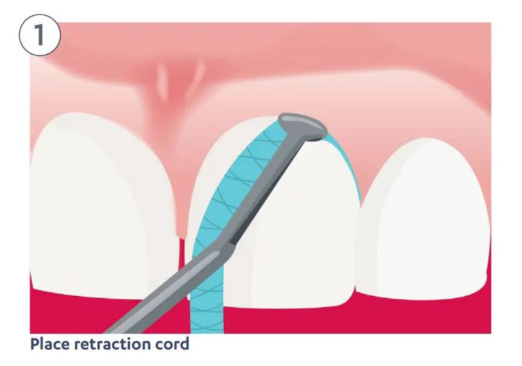
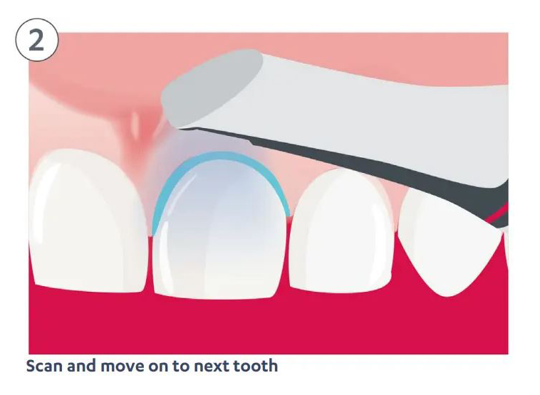
Dr Watt discovered that by placing the retraction cord one tooth at a time, scanning it, and then removing the retraction cord from that tooth and placing it in the next tooth, he was able to avoid the cord displacing the papilla over the margin of the tooth in front.
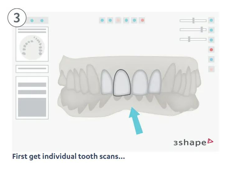
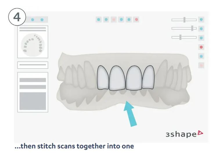
Dr Watt was then able to scan individual tooth preps and stitch the scans together into one scan, noting “the major benefit of using a scanner in this situation is that if there is an area lacking definition, you can simply delete and rescan this area. With a conventional impression, it would have meant replacing some cord and retaking the impression, possibly multiple times.” This would be a nightmare for both the patient and treatment provider.
What the Clinical Accuracy Studies Say
There are many clinical studies available concerning intraoral scanning. The following are a few conclusions from some of them.
“Within the limitations of this study, the zirconia copings fabricated with CAD-CAM using different digitization methods and Ni-Cr copings fabricated using the lost-wax technique and casting produced clinically acceptable marginal and internal discrepancies. No significant differences were found.”
Evaluation of the fit of zirconia copings fabricated by direct and indirect digital impression procedures—Lee B et al, 2017
“In dentate situations, the two tested IOS systems achieved a clinically satisfying accuracy for capturing gingival contour in anterior maxilla, with a comparable or superior precision to the conventional impression. Intraoral digital impressions could be a recommended method to record 3-dimensional gingival contour in the esthetic zone.”
Evaluation of intraoral digital impressions for obtaining gingival contour in the esthetic zone: accuracy outcomes—Wei D, 2020
“Within the limitations of this in vivo study, all of the digital impression systems were capable of measuring quadrant impression with clinically satisfying precision. There are differences in precision between different digital impression systems, but while statistically significant, they all fall within a range which allows the successful production of restorations in the digital workflow.”
In vivo precision of conventional and digital methods for obtaining quadrant dental impressions–Ender A, 2016
In Summary
“The curtain that doctors have been hiding behind, the one that says digital is no good or the quality isn’t there, no longer exists“, says Dr Vincent Prestipino, Prosthodontist.
As seen from these studies, digital impressions deliver equal if not better results when compared with analog impressions. Thankfully there is now extensive clinical research in this area. In this piece, we have given you an introduction and a glimpse of what they say. On the 3Shape website you can find a complete library of clinical studies to review.
Visit our 3Shape blog for more essential insights and know-how from digital dental experts and link them here: https://utm.io/udlqc
Helen Roberts has a PhD in immunology. She has been working for 3Shape since 2018, Her current role is scientific manager, where she works with researchers and universities in addition to maintaining the clinical documentation of 3Shape products and solutions.